Label The Parts Of A Gomphosis
Holbox
Mar 20, 2025 · 6 min read
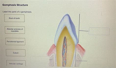
Table of Contents
Labeling the Parts of a Gomphosis: A Deep Dive into Fibrous Joints
Gomphoses, a fascinating type of fibrous joint, are often overlooked in discussions of the skeletal system. Yet, understanding their unique structure and function is crucial to comprehending the mechanics of the skull and the intricate relationship between teeth and bone. This comprehensive guide will delve into the detailed anatomy of a gomphosis, clearly labeling its components and exploring its significance in overall bodily function. We'll also touch upon common misconceptions and provide practical applications of this knowledge.
What is a Gomphosis?
A gomphosis is a fibrous joint characterized by a peg-in-socket articulation. Unlike other fibrous joints like sutures (found in the skull) or syndesmoses (connecting long bones), gomphoses are uniquely specialized for anchoring teeth within the alveolar sockets of the maxilla (upper jaw) and mandible (lower jaw). The word "gomphosis" itself is derived from the Greek word "gomphos," meaning "bolt" or "peg," accurately reflecting the joint's structural nature.
Key Characteristics of a Gomphosis:
-
Fibrous Connective Tissue: The primary component binding the tooth to the socket is a dense, fibrous connective tissue known as the periodontal ligament. This ligament is not merely a passive connector; it plays a vital role in shock absorption, proprioception (sensory feedback regarding tooth position), and maintaining overall tooth stability.
-
Peg-in-Socket Articulation: The "peg" is the root of the tooth, possessing a unique shape that precisely complements the "socket," the alveolar socket within the jawbone. This precise fit is essential for stability and efficient force transmission during chewing.
-
Immobility (Synarthrosis): Gomphoses are classified as synarthroses, meaning they are functionally immobile joints. While there's minimal movement, the periodontal ligament allows for a small degree of physiological movement—necessary for shock absorption and to prevent damage during chewing. This subtle movement is crucial for preventing injury to the tooth and surrounding structures.
Detailed Anatomy: Labeling the Components
Let's meticulously label the crucial parts of a gomphosis, going beyond simple definitions to appreciate the intricacies of this specialized joint:
1. The Tooth:
- Crown: The visible part of the tooth above the gum line. It is covered in enamel, the hardest substance in the human body.
- Neck: The constricted area of the tooth where the crown meets the root.
- Root: The portion of the tooth embedded within the alveolar socket. The root's shape (single, multiple roots) varies depending on the tooth's position and function. The root surface is covered by cementum, a bone-like substance.
- Apical Foramen: A small opening at the tip of the root, through which blood vessels and nerves enter the tooth's pulp cavity. This provides nourishment and sensation to the tooth.
- Pulp Cavity: The central chamber within the tooth containing blood vessels, nerves, and connective tissue (dental pulp).
2. The Alveolar Socket:
- Alveolar Bone (Alveolar Process): The bony socket within the maxilla or mandible that houses the tooth's root. This bone is highly specialized and constantly remodeled in response to forces placed upon the teeth.
- Lamina Dura: A thin layer of compact bone lining the alveolar socket. It provides a smooth, hard surface for the periodontal ligament to attach.
- Bone Marrow: The soft tissue within the alveolar bone containing blood vessels and hematopoietic cells (blood cell precursors).
3. The Periodontal Ligament (PDL):
- Sharpey's Fibers: These are collagen fibers that embed into both the cementum of the tooth root and the lamina dura of the alveolar socket. They are the primary force-transmitting elements within the PDL. They provide strong attachment and are crucial for shock absorption.
- Connective Tissue Cells (Fibroblasts, Osteoblasts, Cementoblasts): These cells are responsible for producing and maintaining the components of the PDL, including collagen fibers, bone, and cementum.
- Blood Vessels and Nerves: The PDL contains a rich network of blood vessels providing nourishment and sensory nerves supplying proprioceptive information.
Functional Significance of Gomphosis:
The gomphosis is not merely a passive structure. Its functional roles are integral to maintaining oral health and overall masticatory function:
-
Force Transmission: During mastication (chewing), the gomphosis efficiently transmits the forces generated by the teeth to the alveolar bone and subsequently to the jawbone. This intricate force distribution prevents damage to individual teeth and the surrounding structures.
-
Shock Absorption: The periodontal ligament acts as a cushion, absorbing the impact of chewing forces. This prevents damage to the tooth root and alveolar bone.
-
Proprioception: Sensory nerve endings within the PDL provide feedback regarding the position and pressure on each tooth. This is crucial for fine motor control during mastication and speaking.
-
Tooth Support and Stability: The tight fit of the tooth within the socket, coupled with the strong attachment of the PDL, provides exceptional support and stability for the teeth, allowing them to withstand the forces of chewing.
Common Misconceptions about Gomphoses:
-
Complete Immobility: While gomphoses are functionally immobile joints, they allow for a small degree of physiological movement. This minor movement is essential for shock absorption and prevention of damage.
-
Simple Structure: The gomphosis is a highly complex structure with multiple components working in harmony to maintain tooth stability and function.
-
Irrelevance in Dentistry: Understanding the anatomy and function of the gomphosis is crucial in dentistry, influencing various procedures such as implant placement, orthodontic treatments, and periodontal disease management.
Practical Applications and Clinical Relevance:
Knowledge of gomphosis anatomy is essential in various dental and medical fields:
-
Orthodontics: Understanding the dynamics of the periodontal ligament is critical for successful orthodontic treatments. Forces applied during orthodontic treatment induce remodeling of the alveolar bone and periodontal ligament, gradually moving teeth into their desired positions.
-
Periodontics: Periodontitis, a common inflammatory disease of the gums and supporting tissues, can lead to damage to the periodontal ligament, compromising tooth stability and potentially resulting in tooth loss. Understanding the structure and function of the gomphosis is crucial in diagnosing and managing periodontal disease.
-
Implantology: The success of dental implants relies heavily on mimicking the natural structure and function of the gomphosis. Careful consideration of the bone density, implant design, and osseointegration (integration of the implant with the surrounding bone) is paramount for successful implant placement.
-
Oral Surgery: Procedures involving tooth extraction or surgical manipulation of the teeth necessitate a thorough understanding of the gomphosis to minimize trauma to the surrounding tissues and promote proper healing.
Conclusion:
The gomphosis, while often overlooked, represents a remarkably intricate and functional joint. Its detailed anatomy, encompassing the tooth, alveolar socket, and periodontal ligament, contributes significantly to mastication, shock absorption, proprioception, and overall oral health. A comprehensive understanding of this specialized joint is essential not only for appreciating the complex mechanics of the skeletal system but also for effective diagnosis, treatment, and management of various dental conditions. The intricate interplay of its components highlights the remarkable design and functionality of the human body. Further research and advancements in understanding the intricacies of the gomphosis will undoubtedly lead to improved dental care and treatment strategies in the future.
Latest Posts
Latest Posts
-
Reasons To Study Operations Management Include
Mar 21, 2025
-
A Disadvantage Of Global Teams For Product Design Is That
Mar 21, 2025
-
Use The Figure Below To Answer The Following Question
Mar 21, 2025
-
Stockholders Have The Right To At Stockholders Meetings
Mar 21, 2025
-
Gaps Or Interruptions In The Myelin Sheath Are Called
Mar 21, 2025
Related Post
Thank you for visiting our website which covers about Label The Parts Of A Gomphosis . We hope the information provided has been useful to you. Feel free to contact us if you have any questions or need further assistance. See you next time and don't miss to bookmark.
