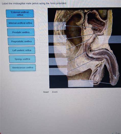
Label The Midsagittal Male Pelvis Using The Hints Provided
Holbox
Apr 01, 2025 · 6 min read

Table of Contents
- Label The Midsagittal Male Pelvis Using The Hints Provided
- Table of Contents
- Labeling the Midsagittal Male Pelvis: A Comprehensive Guide
- I. Bony Structures of the Male Pelvis: A Midsagittal View
- A. The Hip Bone (Os Coxae): A Tripartite Structure
- B. The Sacrum: A Keystone Structure
- C. The Coccyx: A Remnant of the Tail
- II. Ligaments of the Male Pelvis: Crucial for Stability
- A. The Pubic Symphysis: A Fibrocartilaginous Joint
- B. Sacroiliac Joint and Ligaments: Connecting Sacrum and Ilium
- C. Sacrococcygeal Joint: Connecting Sacrum and Coccyx
- III. Other Important Structures in the Midsagittal View
- A. Pelvic Cavity: Enclosing Vital Organs
- B. Sacral Canal and Spinal Cord: A Protective Structure
- C. Pelvic Floor Muscles: Support and Function
- IV. Clinical Significance and Applications
- V. Conclusion: Mastering Pelvic Anatomy
- Latest Posts
- Latest Posts
- Related Post
Labeling the Midsagittal Male Pelvis: A Comprehensive Guide
The male pelvis, a complex structure crucial for weight-bearing, locomotion, and protection of vital organs, presents a fascinating study in anatomy. Understanding its components requires careful examination and precise labeling. This detailed guide provides a thorough walkthrough of labeling a midsagittal section of the male pelvis, using hints to guide you through the process. We will explore the key bony landmarks, ligaments, and associated structures, ensuring a comprehensive understanding of this vital anatomical region.
I. Bony Structures of the Male Pelvis: A Midsagittal View
The midsagittal plane divides the pelvis into two symmetrical halves. Focusing on the midline allows us to identify key structures clearly. The bony pelvis is primarily composed of three fused bones: the two hip bones (ossa coxae) and the sacrum. The coccyx, a small, vestigial bone, is also crucial for understanding the complete pelvic structure.
A. The Hip Bone (Os Coxae): A Tripartite Structure
Each hip bone is formed by the fusion of three separate bones: the ilium, ischium, and pubis. In the midsagittal view, these are readily identifiable.
-
1. Ilium: The largest portion of the hip bone, its superior border forms the iliac crest. The iliac crest, palpable externally, is a crucial landmark for clinicians and anatomists alike. The anterior superior iliac spine (ASIS) and anterior inferior iliac spine (AIIS) are easily identifiable along the anterior border of the ilium. The posterior superior iliac spine (PSIS) and posterior inferior iliac spine (PIIS) are similarly located on the posterior border. In the midsagittal section, you'll see the internal surface of the ilium, including portions of the iliac fossa.
-
2. Ischium: Located inferiorly and posteriorly, the ischium contributes to the acetabulum (the hip socket) and the ischial tuberosity. The ischial spine is a prominent projection visible in the midsagittal view, separating the greater and lesser sciatic notches. The ischial tuberosity, the bony prominence you sit on, is not fully visible in a midsagittal section, but its superior aspect can be seen.
-
3. Pubis: The most anterior portion of the hip bone, the pubis articulates with its counterpart on the opposite side to form the pubic symphysis. The superior and inferior rami of the pubis are clearly visible in a midsagittal view, converging at the pubic symphysis. The pubic tubercle is a small projection located laterally on the superior ramus.
B. The Sacrum: A Keystone Structure
The sacrum, a triangular bone formed by the fusion of five sacral vertebrae, articulates superiorly with the last lumbar vertebra (L5) and inferiorly with the coccyx. In the midsagittal view, you'll see the prominent sacral promontory, which is the anterior edge of the superior articular surface of the first sacral vertebra (S1). The sacral canal, a continuation of the vertebral canal, runs down the midline of the sacrum. The sacral foramina, openings that transmit nerves and blood vessels, are visible on each side. The sacral hiatus, located at the inferior end of the sacrum, is often important clinically for performing caudal epidural anesthesia.
C. The Coccyx: A Remnant of the Tail
The coccyx, the small, triangular bone at the inferior end of the vertebral column, represents the vestigial remains of the tail. In a midsagittal view, its several fused coccygeal vertebrae are usually visible.
II. Ligaments of the Male Pelvis: Crucial for Stability
Several crucial ligaments reinforce the joints of the pelvic girdle, providing stability and supporting the weight of the upper body. A midsagittal view clearly shows some of these essential structures:
A. The Pubic Symphysis: A Fibrocartilaginous Joint
The pubic symphysis, a joint uniting the two pubic bones, is reinforced by the pubic symphysis ligament. This ligament is visible in the midsagittal view, demonstrating the fibrous cartilage that connects the two pubic bones.
B. Sacroiliac Joint and Ligaments: Connecting Sacrum and Ilium
The sacroiliac joints, articulating the sacrum and ilium, are critical for weight transfer. Although a true midsagittal section doesn't fully expose the entire joint surface, you can still visualize the anterior sacroiliac ligament, strengthening the anterior aspect of the joint. The posterior sacroiliac ligaments, stronger and more extensive, are less visible in a midsagittal cut.
C. Sacrococcygeal Joint: Connecting Sacrum and Coccyx
The sacrococcygeal joint connects the sacrum and the coccyx. The sacrococcygeal ligaments, both anterior and posterior, stabilize this articulation, partially visible in a midsagittal view.
III. Other Important Structures in the Midsagittal View
Beyond the bones and ligaments, several other crucial structures are partially or fully visible in a midsagittal section of the male pelvis:
A. Pelvic Cavity: Enclosing Vital Organs
The pelvic cavity, the space enclosed by the bony pelvis, houses vital organs like parts of the urinary system and the rectum. Although internal organs aren't fully depicted in a skeletal preparation, their general location and relation to the pelvic bones can be inferred. The pelvic brim or pelvic inlet, defining the superior limit of the true pelvis, is an important reference point visible in this view. The pelvic outlet, the inferior opening, is also partially visualized.
B. Sacral Canal and Spinal Cord: A Protective Structure
A midsagittal section provides a clear view of the sacral canal which houses the continuation of the spinal cord (the cauda equina). The filum terminale, the fibrous extension of the pia mater, is often visible extending from the conus medullaris.
C. Pelvic Floor Muscles: Support and Function
While the muscles of the pelvic floor are not readily visible on a skeletal midsagittal view, their attachments to bony landmarks are crucial to understanding their function in supporting pelvic organs. Knowing the anatomical location of these muscle attachments (e.g., to the ischial spines, coccyx, and pubic bones) is important for comprehending their contribution to continence and stability.
IV. Clinical Significance and Applications
Understanding the anatomy of the midsagittal male pelvis is crucial in various clinical settings:
- Obstetrics and Gynecology: The pelvic dimensions are vital for assessing the feasibility of vaginal delivery.
- Urology: Knowledge of the pelvic bones and ligaments is critical during urological surgeries.
- Orthopedics: Pelvic fractures often involve the sacrum, ilium, or pubis, necessitating detailed anatomical knowledge for diagnosis and treatment.
- Neurosurgery: Procedures involving the cauda equina require a thorough understanding of the anatomy of the sacral canal.
- Anesthesiology: Caudal epidural anesthesia relies on accurate needle placement into the sacral hiatus.
V. Conclusion: Mastering Pelvic Anatomy
Labeling the midsagittal male pelvis is an exercise in precision and requires a careful understanding of the bony landmarks, ligaments, and associated structures. By systematically approaching the identification and labeling of each component, a thorough comprehension of this complex and clinically relevant anatomical region can be achieved. This detailed guide provides a structured approach, enabling both students and healthcare professionals to enhance their understanding of this essential anatomical structure. Remember, consistent study and practice are key to mastering the intricacies of pelvic anatomy and its clinical applications. Further exploration through anatomical models, atlases, and cadaveric dissection will enhance your knowledge and build a solid foundation for future learning.
Latest Posts
Latest Posts
-
Draw A Reasonable Mechanism For The Following Reaction
Apr 03, 2025
-
Describe What Each Letter Stands For In The Cvp Graph
Apr 03, 2025
-
What Is The Equivalent Resistance Between Points A And B
Apr 03, 2025
-
A Customer Is Traveling To A Branch Office
Apr 03, 2025
-
Setting Up The Math For A Two Step Quantitative Problem
Apr 03, 2025
Related Post
Thank you for visiting our website which covers about Label The Midsagittal Male Pelvis Using The Hints Provided . We hope the information provided has been useful to you. Feel free to contact us if you have any questions or need further assistance. See you next time and don't miss to bookmark.
