Label The Microscopic Anatomy Of Spongy Bone
Holbox
Apr 02, 2025 · 6 min read

Table of Contents
- Label The Microscopic Anatomy Of Spongy Bone
- Table of Contents
- Labeling the Microscopic Anatomy of Spongy Bone: A Comprehensive Guide
- The Unique Structure of Spongy Bone
- Key Microscopic Components to Label:
- Detailed Labeling Exercise: A Step-by-Step Approach
- Clinical Significance of Spongy Bone Anatomy
- Advanced Microscopic Techniques for Spongy Bone Analysis
- Conclusion: Mastering Spongy Bone Microscopic Anatomy
- Latest Posts
- Latest Posts
- Related Post
Labeling the Microscopic Anatomy of Spongy Bone: A Comprehensive Guide
Spongy bone, also known as cancellous bone or trabecular bone, is a type of bone tissue characterized by its porous, honeycomb-like structure. Unlike compact bone, which forms the outer layer of most bones, spongy bone is found inside bones, primarily in the epiphyses (ends) of long bones and within the interior of other bones. Understanding its microscopic anatomy is crucial for comprehending bone function, development, and pathology. This guide provides a detailed walkthrough of labeling the key microscopic features of spongy bone.
The Unique Structure of Spongy Bone
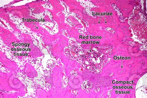
The defining characteristic of spongy bone is its trabecular network. These trabeculae are interconnected bony spicules or plates that create a three-dimensional lattice. This structure is not haphazard; it's highly organized and aligned along lines of stress, maximizing strength with minimal weight. The spaces between the trabeculae are filled with bone marrow, a soft tissue responsible for hematopoiesis (blood cell production) and fat storage.
Key Microscopic Components to Label:
When labeling a microscopic image of spongy bone, you'll encounter several key components. Let's explore each in detail:
-
Trabeculae: These are the thin, bony struts and plates forming the framework of spongy bone. Label these clearly, highlighting their interconnected nature and the varying thickness and orientation. Note how they form a complex network providing significant structural support while remaining lightweight.
-
Bone Marrow: This soft tissue occupies the spaces within the trabecular network. Label the different types of bone marrow present, differentiating between red marrow (hematopoietic, rich in blood cells and progenitor cells) and yellow marrow (primarily composed of fat cells). The proportion of red and yellow marrow changes with age and bone location. Describe the cellularity and overall appearance of the marrow in your labeled diagram.
-
Osteocytes: These are mature bone cells embedded within the bone matrix of the trabeculae. They are housed in small spaces called lacunae. Label both the osteocytes and the lacunae. Mention their crucial role in maintaining bone tissue homeostasis through sensing mechanical stress and regulating bone remodeling.
-
Lacunae: These are small cavities within the bone matrix where osteocytes reside. Label these and connect them to the osteocytes. Note their interconnectedness via canaliculi.
-
Canaliculi: These are tiny canals that radiate from the lacunae, connecting adjacent lacunae and providing pathways for nutrient exchange and communication between osteocytes. Label these delicate channels and emphasize their importance for osteocyte survival and bone tissue health.
-
Osteoblasts: These are bone-forming cells responsible for synthesizing and depositing new bone matrix. They are located on the surfaces of trabeculae. Label these actively building cells and describe their cuboidal or columnar morphology.
-
Osteoclasts: These are large, multinucleated cells responsible for bone resorption (breakdown of bone tissue). They are often found within Howship's lacunae, depressions on the bone surface indicating areas of bone resorption. Label these cells and note their characteristic large size and multiple nuclei.
-
Howship's Lacunae: These are resorption pits or bays on the trabecular surfaces where osteoclasts are actively breaking down bone matrix. Labeling these helps illustrate the dynamic nature of bone remodeling.
Detailed Labeling Exercise: A Step-by-Step Approach
To effectively label a microscopic image of spongy bone, follow these steps:
-
Obtain a Clear Image: Start with a high-quality microscopic image of spongy bone stained appropriately (e.g., hematoxylin and eosin stain). The image should clearly show the trabecular network, bone cells, and marrow spaces.
-
Identify Major Structures: Begin by identifying the major components described above. Look for the interconnected network of trabeculae, the spaces filled with bone marrow, and the presence of osteocytes within lacunae.
-
Label Trabeculae: Clearly label several trabeculae, highlighting their varying thicknesses and orientations. Indicate the bone matrix material within the trabeculae.
-
Label Bone Marrow: Identify and label areas of red and yellow marrow, if present. Note any observable cellular differences between them.
-
Locate and Label Osteocytes: Identify osteocytes within their lacunae and clearly label both structures. Note the presence of canaliculi connecting the lacunae.
-
Identify and Label Osteoblasts and Osteoclasts: Locate and label osteoblasts on the trabecular surfaces. Identify any Howship's lacunae and the osteoclasts within them.
-
Annotate the Image: Add concise labels and annotations to explain the different components and their relationships. Use arrows or lines to connect labels to the specific structures they represent.
-
Add a Title and Legend: Give your labeled image a descriptive title and create a legend explaining the abbreviations or symbols used in your labeling.
Clinical Significance of Spongy Bone Anatomy
Understanding the microscopic anatomy of spongy bone is not merely an academic exercise; it holds significant clinical relevance. Various diseases and conditions directly affect the structure and function of spongy bone:
-
Osteoporosis: This disease is characterized by decreased bone mass and density, leading to weakened trabeculae and increased risk of fractures. Microscopic examination reveals thinned trabeculae with increased porosity and reduced bone matrix.
-
Osteomalacia: This condition results from inadequate mineralization of bone matrix, leading to weakened and softened bones. Microscopic examination reveals poorly mineralized trabeculae with an enlarged osteoid seam (unmineralized bone matrix).
-
Paget's Disease: This chronic bone disorder is characterized by excessive bone remodeling, leading to disorganized and thickened trabeculae. Microscopic examination shows increased osteoclast activity and a mosaic pattern of bone formation.
-
Bone Metastases: Cancer cells can spread to the bone marrow and trabeculae, causing bone destruction and pain. Microscopic examination reveals the presence of malignant cells within the marrow spaces and the erosion of trabeculae.
-
Bone Fractures: Understanding the structural properties of spongy bone helps in analyzing and treating bone fractures, especially those involving the cancellous bone of the epiphyses.
Advanced Microscopic Techniques for Spongy Bone Analysis
Beyond standard light microscopy, various advanced techniques provide more detailed insights into spongy bone structure and composition:
-
Micro-computed tomography (micro-CT): This non-destructive technique generates three-dimensional images of bone microstructure, allowing for quantitative analysis of trabecular bone parameters such as bone volume fraction, trabecular thickness, and connectivity density.
-
Histomorphometry: This method uses quantitative analysis of histological sections to assess bone parameters such as bone formation rate, bone resorption rate, and osteoid thickness.
-
Transmission Electron Microscopy (TEM): This technique provides ultrastructural details of bone cells and the bone matrix, revealing the intricate organization of collagen fibers and mineral crystals.
-
Scanning Electron Microscopy (SEM): SEM imaging provides high-resolution images of bone surfaces, allowing for detailed visualization of trabecular architecture and bone remodeling processes.
Conclusion: Mastering Spongy Bone Microscopic Anatomy
Accurate labeling of the microscopic anatomy of spongy bone requires careful observation, a thorough understanding of its components, and meticulous annotation. This detailed guide provides the necessary knowledge and step-by-step instructions for successfully labeling spongy bone features. By mastering this skill, you gain a deeper appreciation for the complex structure and crucial function of this important bone tissue and its significance in clinical practice and research. Remember that the dynamic nature of bone remodeling, with constant interplay between osteoblasts and osteoclasts, is crucial to understanding the overall health and integrity of spongy bone. The interconnectedness of all the labeled structures emphasizes the systemic nature of bone health. This detailed understanding is essential for comprehending bone-related diseases and developing effective treatments.
Latest Posts
Latest Posts
-
All Of The Following Are Neurotransmitters Except
Apr 05, 2025
-
What Is The Monroes Total Federal Income Tax Withholding
Apr 05, 2025
-
Nerve Fibers From The Medial Aspect Of Each Eye
Apr 05, 2025
-
What Type Of Reaction Involving Stolen Methylamine
Apr 05, 2025
-
Interest Expense On An Interest Bearing Note Is
Apr 05, 2025
Related Post
Thank you for visiting our website which covers about Label The Microscopic Anatomy Of Spongy Bone . We hope the information provided has been useful to you. Feel free to contact us if you have any questions or need further assistance. See you next time and don't miss to bookmark.
