Label The Image Of A Compound Light Microscope
Holbox
Mar 31, 2025 · 6 min read

Table of Contents
- Label The Image Of A Compound Light Microscope
- Table of Contents
- Labeling the Image of a Compound Light Microscope: A Comprehensive Guide
- Key Components of a Compound Light Microscope
- 1. Eyepiece (Ocular Lens):
- 2. Objective Lenses:
- 3. Revolving Nosepiece (Turret):
- 4. Stage:
- 5. Stage Clips:
- 6. Condenser:
- 7. Iris Diaphragm:
- 8. Coarse Adjustment Knob:
- 9. Fine Adjustment Knob:
- 10. Light Source:
- 11. Arm:
- 12. Base:
- 13. Body Tube (Head):
- Advanced Features (Optional Labels):
- Tips for Accurate Labeling:
- Practical Application: Labeling a Microscope Image
- Conclusion: Mastering Microscope Labeling
- Latest Posts
- Latest Posts
- Related Post
Labeling the Image of a Compound Light Microscope: A Comprehensive Guide
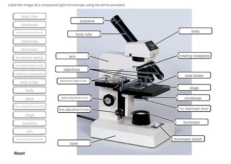
The compound light microscope is a fundamental tool in various scientific disciplines, from biology and microbiology to materials science. Understanding its components and their functions is crucial for effective and accurate use. This comprehensive guide will walk you through the process of labeling a compound light microscope image, explaining the function of each part and providing tips for successful microscopic observation.
Key Components of a Compound Light Microscope
A compound light microscope utilizes a system of lenses to magnify a specimen significantly. Let's explore the critical components you'll need to label:
1. Eyepiece (Ocular Lens):
- Function: The eyepiece is the lens you look through. It typically provides a magnification of 10x. Some microscopes have binocular eyepieces, allowing for comfortable viewing with both eyes.
- Labeling Tip: Clearly identify it as "Eyepiece" or "Ocular Lens".
2. Objective Lenses:
- Function: These are the lenses closest to the specimen. A compound microscope usually has multiple objective lenses with varying magnification powers (e.g., 4x, 10x, 40x, 100x). The 100x lens often requires immersion oil for optimal resolution.
- Labeling Tip: Label each lens with its magnification power (e.g., "4x Objective," "10x Objective," "40x Objective," "100x Objective").
3. Revolving Nosepiece (Turret):
- Function: This rotating structure holds the objective lenses and allows you to easily switch between different magnification levels.
- Labeling Tip: Simply label it as "Revolving Nosepiece" or "Turret".
4. Stage:
- Function: The flat platform where you place the microscope slide containing your specimen. Many stages have stage clips to hold the slide securely in place. Some advanced models feature mechanical stages for precise movement.
- Labeling Tip: Label it as "Stage". If it has clips, you might add "Stage Clips".
5. Stage Clips:
- Function: These metal clips hold the microscope slide firmly on the stage, preventing it from shifting during observation.
- Labeling Tip: Label them as "Stage Clips".
6. Condenser:
- Function: The condenser focuses light from the light source onto the specimen. It improves the clarity and resolution of the image by controlling the light cone. It often has a diaphragm to adjust the amount of light passing through.
- Labeling Tip: Label it as "Condenser".
7. Iris Diaphragm:
- Function: Located within the condenser, the iris diaphragm controls the amount of light passing through the condenser, affecting contrast and resolution. Adjusting this is crucial for optimal viewing.
- Labeling Tip: Label it as "Iris Diaphragm" or "Condenser Diaphragm".
8. Coarse Adjustment Knob:
- Function: This large knob allows for rapid focusing of the specimen by moving the stage up and down. It is primarily used with lower magnification objectives.
- Labeling Tip: Label it as "Coarse Adjustment Knob".
9. Fine Adjustment Knob:
- Function: This smaller knob provides precise focusing of the specimen, especially crucial at higher magnifications. It makes fine adjustments to the stage's vertical position.
- Labeling Tip: Label it as "Fine Adjustment Knob".
10. Light Source:
- Function: This provides illumination for viewing the specimen. Modern microscopes usually have built-in LED light sources. Older models might use a mirror to reflect external light.
- Labeling Tip: Label it as "Light Source" or "Illuminator".
11. Arm:
- Function: The vertical structure that connects the base to the body tube and supports the eyepiece and objective lenses. It's used to carry the microscope.
- Labeling Tip: Label it as "Arm".
12. Base:
- Function: The bottom support structure of the microscope. It provides stability and houses the light source in many models.
- Labeling Tip: Label it as "Base".
13. Body Tube (Head):
- Function: The tube connecting the eyepiece to the objective lenses. It maintains the proper alignment between the lenses.
- Labeling Tip: Label it as "Body Tube" or "Head".
Advanced Features (Optional Labels):
Some advanced compound light microscopes might include additional features requiring labeling:
- Mechanical Stage: Allows for precise x-y movement of the slide. Label as "Mechanical Stage".
- Köhler Illumination Controls: Allows for optimized illumination for even brightness and contrast across the field of view. Label the individual components appropriately (e.g., "Field Diaphragm," "Condenser Adjustment Knob").
- Phase Contrast Components: For enhancing the contrast of transparent specimens. Label the elements specific to phase contrast (e.g., "Phase Ring," "Annulus").
- Fluorescence Illumination System: For viewing fluorescently labeled specimens. Label the specific parts (e.g., "Excitation Filter," "Emission Filter").
Tips for Accurate Labeling:
- Use clear and concise labels: Avoid ambiguity. Use standard terminology.
- Use a consistent font and size: Maintain visual uniformity.
- Ensure labels are easy to read: Avoid overlapping labels and cluttered diagrams.
- Use arrows to point to the labeled components: This helps readers directly associate the label with the corresponding part of the microscope.
- Consider using different colors for different components: This aids in visual organization.
- Include a legend if necessary: This is especially useful if you have many components to label.
Practical Application: Labeling a Microscope Image
Let's apply these labeling principles to a hypothetical scenario. Imagine you have an image of a compound light microscope. Follow these steps:
- Identify each part: Carefully examine the image and identify each component based on its visual characteristics and location.
- Create a list of components: Make a list of all the parts you've identified.
- Label each part clearly: Add labels to the image using clear and concise terminology, possibly with arrows pointing to the specific parts. Use the specific names from the previous sections of this guide.
- Check for accuracy: Verify that all the labels are correct and that the image is easy to understand.
- Consider adding a title and caption: This will provide context and information.
For example, your labeled image might look something like this (replace with actual image):
[Image of Compound Light Microscope]
- Eyepiece (Ocular Lens)
- Revolving Nosepiece (Turret)
- 4x Objective Lens
- 10x Objective Lens
- 40x Objective Lens
- 100x Objective Lens
- Stage
- Stage Clips
- Condenser
- Iris Diaphragm
- Coarse Adjustment Knob
- Fine Adjustment Knob
- Light Source
- Arm
- Base
- Body Tube
Conclusion: Mastering Microscope Labeling
Mastering the art of labeling a compound light microscope image is a crucial skill for anyone working with this essential scientific instrument. By understanding the function of each component and following the tips outlined in this guide, you can create accurate, clear, and informative labeled diagrams that aid in your understanding and communication of microscopic techniques and observations. This understanding is crucial not only for academic settings but also for various professional fields relying on microscopy. Remember that clear communication through accurate labeling is essential for effective scientific practice.
Latest Posts
Latest Posts
-
In Marketing An Exchange Refers To
Apr 03, 2025
-
Identify The Atom With The Following Ground State Electron Configuration
Apr 03, 2025
-
Benicio Del Toro Es Un Beisbolista Puertorrique A O
Apr 03, 2025
-
Which Statement Describes A Clients Tidal Volume
Apr 03, 2025
-
For Each Of The Following Compute The Present Value
Apr 03, 2025
Related Post
Thank you for visiting our website which covers about Label The Image Of A Compound Light Microscope . We hope the information provided has been useful to you. Feel free to contact us if you have any questions or need further assistance. See you next time and don't miss to bookmark.
