Lab Report 9 Bacterial Flagella And Motility Testing
Holbox
Mar 13, 2025 · 6 min read
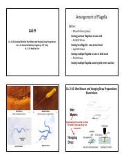
Table of Contents
- Lab Report 9 Bacterial Flagella And Motility Testing
- Table of Contents
- Lab Report 9: Bacterial Flagella and Motility Testing
- Understanding Bacterial Flagella and Motility
- Types of Flagellar Arrangements:
- The Structure and Function of Bacterial Flagella:
- Methods for Detecting Bacterial Motility
- 1. Wet Mount Microscopy:
- 2. Semi-Solid Agar Stab Inoculation:
- Procedure: A Step-by-Step Guide
- Materials:
- Procedure: Wet Mount Microscopy
- Procedure: Semi-Solid Agar Stab Inoculation
- Results and Interpretation
- Data Presentation:
- Discussion and Conclusion
- Key Discussion Points:
- References
- Further Considerations
- Latest Posts
- Latest Posts
- Related Post
Lab Report 9: Bacterial Flagella and Motility Testing
This comprehensive guide delves into the intricacies of bacterial flagella and motility testing, providing a detailed framework for writing a compelling and informative lab report. We'll cover the theoretical background, practical procedures, results interpretation, and crucial elements for effective scientific communication. This report aims to equip you with the knowledge and skills necessary to confidently analyze and present your findings on bacterial motility.
Understanding Bacterial Flagella and Motility
Bacterial motility, the ability of bacteria to move independently, is a critical characteristic influencing their survival, pathogenesis, and ecological roles. This movement is primarily facilitated by flagella, long, whip-like appendages extending from the bacterial cell surface. Different bacterial species exhibit diverse flagellar arrangements, which are key identifiers in bacterial classification and identification.
Types of Flagellar Arrangements:
- Monotrichous: A single flagellum at one pole of the cell.
- Amphitrichous: A single flagellum at each pole of the cell.
- Lophotrichous: A cluster of flagella at one or both poles of the cell.
- Peritrichous: Flagella distributed over the entire cell surface.
The arrangement and number of flagella significantly impact the bacterium's motility pattern. For instance, peritrichous flagella often result in a tumbling or swarming movement, while monotrichous flagella lead to more directed movement.
The Structure and Function of Bacterial Flagella:
Bacterial flagella are complex molecular machines composed of several key components:
- Filament: The long, helical structure composed of the protein flagellin.
- Hook: A curved segment connecting the filament to the basal body.
- Basal Body: A complex structure embedded in the cell membrane and cell wall, responsible for rotation of the flagellum. The basal body contains a motor that utilizes the proton motive force (PMF) to drive flagellar rotation.
Understanding the structure and function of flagella is essential for interpreting motility test results. Any disruption to these components will directly affect the bacterium's ability to move.
Methods for Detecting Bacterial Motility
Several methods exist for assessing bacterial motility. This section will focus on two common techniques:
1. Wet Mount Microscopy:
This simple technique allows direct observation of bacterial motility under a light microscope. A small sample of bacterial culture is placed on a clean glass slide, covered with a coverslip, and observed for movement. While this method provides a direct visual assessment, it's relatively less precise than other techniques and can be challenging to differentiate true motility from Brownian motion (random movement due to collisions with water molecules).
2. Semi-Solid Agar Stab Inoculation:
This method employs a semi-solid agar medium (lower agar concentration compared to solid agar), allowing motile bacteria to swim throughout the medium. The bacterium is inoculated by stabbing the agar with an inoculating needle. Motile bacteria will exhibit growth radiating outward from the inoculation line, creating a cloudy or fuzzy appearance throughout the tube. Non-motile bacteria will only grow along the inoculation line, forming a distinct, localized growth pattern. This method is widely considered more reliable than wet mount microscopy for determining bacterial motility.
Procedure: A Step-by-Step Guide
This section details the standard procedure for performing bacterial flagella and motility testing using both wet mount microscopy and semi-solid agar stab inoculation.
Materials:
- Bacterial cultures (various species)
- Microscope slides and coverslips
- Inoculating loops and needles
- Sterile distilled water
- Semi-solid agar tubes
- Light microscope
- Incubator
Procedure: Wet Mount Microscopy
- Prepare a Bacterial Suspension: Aseptically transfer a small amount of bacterial culture into a drop of sterile distilled water on a clean microscope slide.
- Apply Coverslip: Carefully place a coverslip over the suspension, avoiding air bubbles.
- Observe under Microscope: Observe the slide under low and high power magnification. Look for directional movement of the bacteria. Differentiate true motility from Brownian motion. Note the motility pattern (e.g., tumbling, gliding, swimming).
- Record Observations: Carefully document your observations, including the bacterial species, motility pattern, and any other relevant observations.
Procedure: Semi-Solid Agar Stab Inoculation
- Inoculate Semi-Solid Agar: Using a sterile inoculating needle, carefully stab the semi-solid agar tube straight down to the bottom, then withdraw along the same path, avoiding excessive movement.
- Incubate: Incubate the tube at the optimal temperature for the bacterial species for 24-48 hours.
- Observe Growth Pattern: After incubation, observe the growth pattern in the semi-solid agar. Motile bacteria will show growth radiating outward from the inoculation line. Non-motile bacteria will only show growth along the inoculation line.
- Record Observations: Document your observations, noting the bacterial species and the growth pattern (motile or non-motile).
Results and Interpretation
The results obtained from both methods should be carefully analyzed and compared. Discrepancies might arise due to factors such as the age of the culture or variations in experimental conditions. Always correlate your findings from both methods.
Data Presentation:
- Table: A well-organized table summarizing the bacterial species, flagellar arrangement (if known), motility observation from wet mount microscopy, and motility observation from semi-solid agar stab inoculation.
- Figures: Include micrographs from the wet mount microscopy and photographs of the semi-solid agar stab cultures illustrating the growth patterns.
- Detailed descriptions: Provide a comprehensive narrative description of your observations, detailing the motility patterns observed and correlating them with the bacterial species' known flagellar arrangement.
Discussion and Conclusion
The discussion section is critical for demonstrating your understanding of the experimental results and their implications.
Key Discussion Points:
- Correlation of Results: Discuss the correlation between your observations from both testing methods. Explain any discrepancies and offer potential explanations.
- Relationship between Flagellar Arrangement and Motility: Relate your observations to the known flagellar arrangements of the bacterial species tested. Discuss how the flagellar arrangement influences the motility pattern.
- Limitations of the Methods: Acknowledge any limitations of the methods used, such as the potential for misinterpreting Brownian motion as true motility in wet mount microscopy.
- Relevance to Bacterial Pathogenesis: Discuss the significance of bacterial motility in the context of bacterial pathogenesis and infection. Motility can enhance bacterial colonization, invasion, and evasion of the host immune system.
- Clinical Significance: Discuss the clinical relevance of accurately identifying bacterial motility in disease diagnosis and treatment. The presence or absence of motility can be a crucial factor in identifying pathogenic bacteria.
The conclusion should summarize your key findings and reiterate the importance of accurate bacterial motility testing in microbiology. This section should highlight the overall significance of your findings and their contributions to understanding bacterial biology.
References
This section should list all cited sources using a consistent citation style (e.g., APA, MLA).
Further Considerations
- Additional Motility Tests: Explore other motility testing methods such as the hanging drop technique or the use of specialized motility media.
- Advanced Microscopy Techniques: Consider using phase-contrast microscopy or dark-field microscopy for enhanced visualization of bacterial motility.
- Molecular Techniques: Explore advanced molecular techniques for studying flagellar genes and their role in motility.
By meticulously following these guidelines and incorporating detailed explanations, your lab report on bacterial flagella and motility testing will be thorough, informative, and effectively communicate your scientific findings. Remember to always prioritize accuracy, clarity, and adherence to scientific principles.
Latest Posts
Latest Posts
-
How Many Kg In 19 Stone
May 21, 2025
-
How Far Is 200km In Miles
May 21, 2025
-
How Much Is 58kg In Stone
May 21, 2025
-
How Much Is 19 Stone In Kg
May 21, 2025
-
How Many Minutes Are In 11 Hours
May 21, 2025
Related Post
Thank you for visiting our website which covers about Lab Report 9 Bacterial Flagella And Motility Testing . We hope the information provided has been useful to you. Feel free to contact us if you have any questions or need further assistance. See you next time and don't miss to bookmark.