Identify The Following Cell Type And Label Its Three Parts
Holbox
Mar 28, 2025 · 7 min read
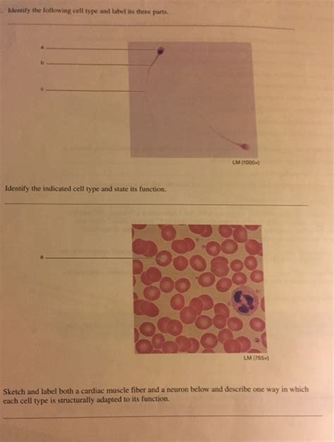
Table of Contents
- Identify The Following Cell Type And Label Its Three Parts
- Table of Contents
- Identify the Following Cell Type and Label Its Three Parts: A Deep Dive into Neuron Structure and Function
- Identifying the Neuron: The Master Communicator
- The Three Primary Parts of a Neuron: A Functional Trinity
- 1. Soma (Cell Body): The Neuron's Control Center
- 2. Dendrites: The Receiving Antennas
- 3. Axon: The Transmission Cable
- Beyond the Basics: Neuron Diversity and Functional Specialization
- Conclusion: A Foundation for Further Exploration
- Latest Posts
- Latest Posts
- Related Post
Identify the Following Cell Type and Label Its Three Parts: A Deep Dive into Neuron Structure and Function
The human body is a marvel of intricate biological engineering, a complex tapestry woven from trillions of cells. Among these, neurons stand out as the fundamental units of the nervous system, responsible for receiving, processing, and transmitting information throughout the body. Understanding the structure of a neuron is crucial to comprehending how our brains, spinal cords, and peripheral nerves function. This article will delve into the identification of a neuron, labeling its three primary parts, and exploring their individual roles in the intricate process of neural communication.
Identifying the Neuron: The Master Communicator
Neurons are highly specialized cells characterized by their unique morphology and function. Unlike many other cell types that primarily focus on structural support or metabolic processes, neurons are dedicated to rapid communication. This specialized function dictates their distinctive structure. They possess elongated processes, extending outwards from the main cell body, enabling them to receive signals from other neurons and transmit signals to their targets.
Several key features distinguish neurons from other cells:
- Long Processes: Neurons possess extensive branching processes extending from the cell body, allowing for communication across significant distances within the nervous system.
- Synaptic Connections: Neurons communicate with each other and with target cells (like muscle cells or gland cells) at specialized junctions called synapses. These junctions allow for the transmission of signals via neurotransmitters.
- Excitable Membranes: The neuron's cell membrane is highly excitable, meaning it can rapidly change its electrical potential, generating and propagating electrical signals (action potentials) that are the basis of neural communication.
- Specialized organelles: Neurons contain a unique complement of organelles tailored to support their high energy demands and the precise mechanisms of neurotransmission.
The image accompanying this article would ideally show a clearly labeled neuron, highlighting the three primary parts discussed below. Without a specific image to reference, the following description will focus on generalized neuron structure.
The Three Primary Parts of a Neuron: A Functional Trinity
A neuron is classically divided into three main parts:
1. Soma (Cell Body): The Neuron's Control Center
The soma, or cell body, is the neuron's central hub. It contains the nucleus, the powerhouse of the cell, holding the genetic material (DNA) responsible for the neuron's structure and function. The soma is also responsible for:
- Protein Synthesis: The soma houses ribosomes and the endoplasmic reticulum, crucial components for protein synthesis. These proteins are essential for the neuron's structure, function, and neurotransmission. Many of these proteins are transported to other parts of the neuron.
- Metabolic Processes: The soma carries out vital metabolic functions, providing energy for the neuron's activities. This includes generating ATP (adenosine triphosphate), the cell's primary energy currency.
- Integration of Signals: The soma receives signals from the dendrites and integrates them. It's the decision-making center, determining whether to trigger an action potential.
The structure of the soma is critical. Its size and shape vary depending on the neuron type, impacting its ability to integrate signals and communicate effectively. The extensive cytoskeleton within the soma contributes to its shape and helps in maintaining the cell's structure. It also plays a key role in transporting molecules to and from the other parts of the neuron.
2. Dendrites: The Receiving Antennas
Dendrites are branching extensions of the soma, acting as the neuron's primary receivers of incoming signals. These highly branched structures increase the surface area available for receiving input from other neurons. Their arborization (branching pattern) is incredibly diverse, with variations reflecting the neuron's role in the neural circuit.
- Synaptic Inputs: Dendrites receive signals from other neurons at specialized junctions called synapses. These synapses can be excitatory, promoting action potentials, or inhibitory, suppressing them.
- Signal Integration: Dendrites don't simply passively receive signals; they actively process them. The summation of excitatory and inhibitory signals determines whether the neuron will ultimately fire an action potential.
- Spines: Many dendrites are studded with small protrusions called dendritic spines. These spines further increase the surface area for synaptic inputs and play a crucial role in synaptic plasticity, the ability of synapses to strengthen or weaken over time. This is fundamental to learning and memory.
The complexity of dendritic branching directly impacts the neuron's integrative capacity. Neurons with many branches can integrate signals from a large number of other neurons, significantly impacting the processing of information within the nervous system. The density and distribution of spines on dendrites also contribute to the neuron's computational capabilities.
3. Axon: The Transmission Cable
The axon is a long, slender projection extending from the soma. It's the neuron's transmission cable, responsible for carrying electrical signals (action potentials) away from the soma to other neurons or target cells. The axon's structure is crucial for its function:
- Myelin Sheath: Many axons are insulated by a myelin sheath, a fatty substance produced by glial cells (oligodendrocytes in the central nervous system and Schwann cells in the peripheral nervous system). The myelin sheath dramatically increases the speed of action potential propagation, ensuring rapid communication.
- Nodes of Ranvier: The myelin sheath is not continuous; it's interrupted by gaps called Nodes of Ranvier. Action potentials "jump" between these nodes (saltatory conduction), further accelerating signal transmission.
- Axon Terminals: At its end, the axon branches into numerous axon terminals, each forming a synapse with another neuron or target cell. These terminals contain neurotransmitters, chemicals that transmit signals across the synapse.
The length and diameter of the axon vary widely, influencing the speed and efficiency of signal transmission. Longer axons allow for communication over greater distances within the nervous system. A larger axon diameter also promotes faster conduction speeds. The health and integrity of the myelin sheath are crucial for efficient signal transmission; damage to myelin, as seen in multiple sclerosis, can significantly impair neural function.
Beyond the Basics: Neuron Diversity and Functional Specialization
While the three primary parts – soma, dendrites, and axon – are common to all neurons, there is significant diversity in their morphology and function. Neurons are not all created equal; their structure is finely tuned to their specific role within the nervous system.
For instance:
- Sensory neurons: These neurons transmit information from sensory receptors (e.g., in the skin, eyes, ears) to the central nervous system. They often have long axons and specialized dendrites optimized for receiving specific sensory information.
- Motor neurons: These neurons transmit signals from the central nervous system to muscles or glands, causing them to contract or secrete. They typically have long axons that extend to their target tissues.
- Interneurons: These neurons connect sensory and motor neurons within the central nervous system. They are highly diverse in their morphology and play a crucial role in processing information.
The specific arrangement of dendrites, the length and myelination of axons, and the type of neurotransmitters released at synapses all contribute to the functional specialization of different neuron types. Understanding this diversity is critical for understanding the complexity of neural circuits and the intricate workings of the brain.
Conclusion: A Foundation for Further Exploration
This article has provided a foundational understanding of neuron structure, identifying the three primary parts – soma, dendrites, and axon – and highlighting their crucial roles in neural communication. The intricate interplay between these parts, along with the vast diversity of neuron types, underlies the complexity and sophistication of the nervous system. Further exploration into the specific mechanisms of neurotransmission, synaptic plasticity, and neural circuit formation will reveal even greater insights into the fascinating world of neuroscience. Understanding the basic structure of the neuron is a crucial first step towards comprehending the complexities of the human brain and the wonders of neural function. The field continues to expand with ongoing research into the microscopic world that underpins our thoughts, feelings, and actions.
Latest Posts
Latest Posts
-
Art Labeling Activity Functions Of Antibodies
Apr 01, 2025
-
Select The Correct Iupac Name For Each Unsaturated Hydrocarbon
Apr 01, 2025
-
On An Angle Tackle The Defender Tracks
Apr 01, 2025
-
Iso Certification Is Similar To The Baldrige Award
Apr 01, 2025
-
Which Of The Following Is A Function Of A Protein
Apr 01, 2025
Related Post
Thank you for visiting our website which covers about Identify The Following Cell Type And Label Its Three Parts . We hope the information provided has been useful to you. Feel free to contact us if you have any questions or need further assistance. See you next time and don't miss to bookmark.
