Drag The Appropriate Labels To Their Respective Targets. Facial Nerve
Holbox
Mar 21, 2025 · 6 min read
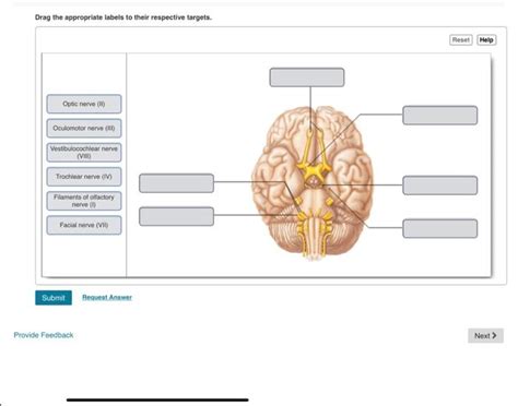
Table of Contents
- Drag The Appropriate Labels To Their Respective Targets. Facial Nerve
- Table of Contents
- Drag the Appropriate Labels to Their Respective Targets: A Comprehensive Guide to the Facial Nerve
- I. Origin and Course of the Facial Nerve: A Journey Through the Cranial Vault
- 1. Internal Acoustic Meatus (IAM): The First Stop
- 2. Facial Canal: A Winding Path
- 3. Geniculate Ganglion: A Sensory Relay
- 4. Branches Within the Temporal Bone: Beyond the Geniculate Ganglion
- 5. Stylomastoid Foramen: Exit from the Temporal Bone
- II. Extracranial Course and Branches: Reaching the Target Muscles
- 1. Temporal Branch: The Upper Face
- 2. Zygomatic Branch: The Cheek Region
- 3. Buccal Branch: The Cheek and Mouth Region
- 4. Marginal Mandibular Branch: The Lower Lip and Chin
- 5. Cervical Branch: The Neck Region
- III. Clinical Significance: Understanding Facial Nerve Palsy
- 1. Causes of Facial Nerve Palsy
- 2. Symptoms of Facial Nerve Palsy
- 3. Diagnosis and Treatment
- IV. Interactive Learning: Reinforcing Your Knowledge
- V. Conclusion: Mastering the Intricacies of the Facial Nerve
- Latest Posts
- Latest Posts
- Related Post
Drag the Appropriate Labels to Their Respective Targets: A Comprehensive Guide to the Facial Nerve
The facial nerve (CN VII) is a complex cranial nerve with a fascinating and intricate anatomy. Understanding its course, branches, and functions is crucial for anyone studying anatomy, neurology, or related fields. This article provides a comprehensive overview of the facial nerve, using an interactive "drag-and-drop" approach to reinforce learning and improve comprehension. While we can't actually implement a drag-and-drop interface in this text format, we will utilize bold headings, bullet points, and detailed descriptions to mimic the experience and help you master the anatomy of the facial nerve.
I. Origin and Course of the Facial Nerve: A Journey Through the Cranial Vault
The facial nerve's journey begins within the brainstem, specifically at the pons. From its origin, it undertakes a complex course, passing through several crucial structures before reaching its peripheral targets.
1. Internal Acoustic Meatus (IAM): The First Stop
Leaving the brainstem, the facial nerve enters the internal acoustic meatus (IAM), a bony canal within the temporal bone. Here, it lies alongside the vestibulocochlear nerve (CN VIII), sharing this pathway before diverging.
2. Facial Canal: A Winding Path
The facial nerve continues its journey within the facial canal, a tortuous passage through the temporal bone. This canal is not a straight shot; it involves several bends and turns, making its precise anatomy challenging but fascinating to study.
3. Geniculate Ganglion: A Sensory Relay
Along the facial canal, the facial nerve encounters the geniculate ganglion, a crucial sensory ganglion that houses cell bodies of the nerve's sensory fibers. These fibers receive sensory input from the taste buds of the anterior two-thirds of the tongue and the ear canal. The geniculate ganglion also marks a significant branching point.
4. Branches Within the Temporal Bone: Beyond the Geniculate Ganglion
Several important branches arise from the facial nerve within the temporal bone:
-
Greater Petrosal Nerve: This branch carries parasympathetic fibers to the lacrimal gland (tear production), nasal mucosa, and salivary glands (e.g., submandibular and sublingual glands). Its role in tear production is critical to understanding dry eye conditions.
-
Stapedius Nerve: This branch innervates the stapedius muscle, a tiny muscle in the middle ear responsible for dampening loud sounds. Damage to this nerve can result in hyperacusis (increased sensitivity to sound).
5. Stylomastoid Foramen: Exit from the Temporal Bone
After traversing the facial canal, the facial nerve finally exits the temporal bone through the stylomastoid foramen. This marks a crucial transition point as the nerve enters the parotid gland.
II. Extracranial Course and Branches: Reaching the Target Muscles
Upon exiting the stylomastoid foramen, the facial nerve enters the parotid gland, a large salivary gland located in front of the ear. Within the parotid gland, the facial nerve divides into its five major terminal branches. Understanding the innervation pattern of these branches is essential for diagnosing facial nerve palsy.
1. Temporal Branch: The Upper Face
The temporal branch innervates the frontalis muscle (responsible for raising the eyebrows), orbicularis oculi muscle (closing the eyes), and corrugator supercilii muscle (frowning). Damage to this branch results in difficulty in raising the eyebrows and closing the eyes completely.
2. Zygomatic Branch: The Cheek Region
The zygomatic branch innervates the muscles responsible for raising the corners of the mouth and smiling: the zygomaticus major and minor muscles. Weakness in this branch leads to a flattened smile.
3. Buccal Branch: The Cheek and Mouth Region
The buccal branch innervates the buccinator muscle (cheek muscle) and the muscles around the mouth, playing a significant role in lip movements, chewing, and whistling. Damage here results in difficulty in puffing out the cheeks and controlling lip movements.
4. Marginal Mandibular Branch: The Lower Lip and Chin
The marginal mandibular branch innervates the muscles of the lower lip and chin, particularly the depressor anguli oris (pulling down the corners of the mouth) and the mentalis muscle (raising the chin). Weakness here leads to drooping of the lower lip and difficulty in controlling lower lip movements.
5. Cervical Branch: The Neck Region
The cervical branch innervates the platysma muscle, a superficial muscle in the neck. This muscle helps in lowering the mandible and expressing emotions. Damage to this branch causes weakness in this region.
III. Clinical Significance: Understanding Facial Nerve Palsy
Damage to the facial nerve, commonly known as facial nerve palsy or Bell's palsy, can have significant consequences depending on the location and severity of the injury.
1. Causes of Facial Nerve Palsy
Facial nerve palsy can arise from various causes, including:
- Idiopathic (Bell's Palsy): This is the most common cause, with an unknown etiology often attributed to viral infections.
- Trauma: Injury to the facial nerve during surgery or from other trauma.
- Tumors: Tumors compressing or infiltrating the facial nerve.
- Infections: Infections affecting the nerve itself or surrounding structures.
- Stroke: Damage to the brain affecting the facial nerve nuclei.
2. Symptoms of Facial Nerve Palsy
The symptoms of facial nerve palsy vary depending on the location and extent of the damage:
- Facial Weakness or Paralysis: This is the hallmark symptom, affecting one side of the face.
- Drooping of the Mouth: Difficulty in controlling lip movements and smiling.
- Difficulty Closing the Eye: Inability to fully close the affected eye.
- Dry Eye: Reduced tear production due to damage to the greater petrosal nerve.
- Hyperacusis: Increased sensitivity to sound due to damage to the stapedius nerve.
- Loss of Taste: Affected taste sensation in the anterior two-thirds of the tongue.
3. Diagnosis and Treatment
Diagnosis involves a thorough neurological examination, including testing the facial muscles and evaluating for other neurological deficits. Treatment depends on the underlying cause and may include:
- Steroids: To reduce inflammation.
- Antivirals: If a viral infection is suspected.
- Surgical Intervention: In cases of severe damage or compression.
- Physical Therapy: To help restore facial muscle function.
IV. Interactive Learning: Reinforcing Your Knowledge
While a true drag-and-drop interface isn't possible here, let's use a descriptive approach to solidify your understanding. Imagine you have to drag labels representing the various branches and structures onto a diagram of the facial nerve. Using the information above, mentally "drag" each label to its correct location.
For example:
- Internal Acoustic Meatus: You would mentally drag this label to the point where the facial nerve enters the temporal bone.
- Geniculate Ganglion: You would place this label along the facial canal where the sensory ganglion is located.
- Stylomastoid Foramen: This label goes to the point where the nerve exits the temporal bone.
- Temporal Branch: This label is located in the upper face.
- Zygomatic Branch: This label would be placed on the cheek region.
- Buccal Branch: This label would go near the cheek and mouth.
- Marginal Mandibular Branch: This is positioned on the lower lip and chin.
- Cervical Branch: Place this label on the neck.
By mentally performing this exercise, you can strengthen your grasp of the facial nerve's anatomy.
V. Conclusion: Mastering the Intricacies of the Facial Nerve
The facial nerve is a marvel of anatomical engineering, with a complex course and diverse functions. Understanding its anatomy and clinical significance is paramount for healthcare professionals and students alike. By reviewing the detailed descriptions and engaging in the mental "drag-and-drop" exercise, you can build a solid foundation of knowledge regarding this important cranial nerve. Remember to consult reliable anatomical resources and texts for further detailed study. The information provided here serves as a comprehensive overview, but further exploration will enhance your understanding of this complex yet fascinating anatomical structure. Continuing your learning journey and delving deeper into the specifics will enable you to master the intricacies of the facial nerve and its clinical implications. Good luck!
Latest Posts
Latest Posts
-
Which Of The Following Is True Regarding Chimpanzee Hunting
Mar 28, 2025
-
Which Component In The Accompanying Figure Is Cholesterol
Mar 28, 2025
-
A Companys Values Or Core Values Concern
Mar 28, 2025
-
Refer To Figure 4 17 At A Price Of
Mar 28, 2025
-
A Companys Ethical Code Of Conduct Is Not
Mar 28, 2025
Related Post
Thank you for visiting our website which covers about Drag The Appropriate Labels To Their Respective Targets. Facial Nerve . We hope the information provided has been useful to you. Feel free to contact us if you have any questions or need further assistance. See you next time and don't miss to bookmark.
