Correctly Label The Following Internal Anatomy Of The Heart.
Holbox
Apr 01, 2025 · 6 min read
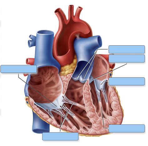
Table of Contents
- Correctly Label The Following Internal Anatomy Of The Heart.
- Table of Contents
- Correctly Label the Following Internal Anatomy of the Heart: A Comprehensive Guide
- The Four Chambers: The Heart's Working Units
- 1. The Right Atrium:
- 2. The Right Ventricle:
- 3. The Left Atrium:
- 4. The Left Ventricle:
- The Heart Valves: Ensuring Unidirectional Blood Flow
- 1. Atrioventricular Valves (AV Valves):
- 2. Semilunar Valves:
- The Conduction System: Orchestrating the Heartbeat
- 1. Sinoatrial (SA) Node:
- 2. Atrioventricular (AV) Node:
- 3. Bundle of His (Atrioventricular Bundle):
- 4. Bundle Branches:
- 5. Purkinje Fibers:
- The Coronary Arteries: Nourishing the Heart Muscle
- 1. Left Coronary Artery:
- 2. Right Coronary Artery:
- Beyond the Basic Structures: Further Exploration
- Latest Posts
- Latest Posts
- Related Post
Correctly Label the Following Internal Anatomy of the Heart: A Comprehensive Guide
The human heart, a remarkable organ, tirelessly pumps blood throughout our bodies, sustaining life itself. Understanding its intricate internal anatomy is crucial for appreciating its function and the complexities of the cardiovascular system. This comprehensive guide will delve into the internal structures of the heart, providing detailed descriptions and visual aids to help you correctly label each component.
The Four Chambers: The Heart's Working Units
The heart is divided into four chambers: two atria (singular: atrium) and two ventricles. These chambers work in a coordinated sequence to ensure efficient blood circulation.
1. The Right Atrium:
The right atrium receives deoxygenated blood returning from the body through the superior vena cava (carrying blood from the upper body) and the inferior vena cava (carrying blood from the lower body). It also receives blood from the coronary sinus, which drains blood from the heart muscle itself. Notice the tricuspid valve, situated between the right atrium and the right ventricle. This valve prevents backflow of blood into the atrium when the ventricle contracts. The right atrium also possesses a small appendage called the right auricle, an extension that slightly increases its volume. The right atrium’s muscular walls are relatively thin, reflecting its lower pressure compared to the left atrium.
2. The Right Ventricle:
The right ventricle receives deoxygenated blood from the right atrium via the tricuspid valve. Its primary function is to pump this blood to the lungs for oxygenation. The pulmonary valve controls the outflow of blood from the right ventricle into the pulmonary artery. The right ventricle's walls are thicker than the right atrium's, but significantly thinner than the left ventricle's, reflecting the lower pressure required to pump blood to the nearby lungs. The internal surface of the right ventricle displays characteristic trabeculae carneae, muscular ridges that enhance ventricular contraction.
3. The Left Atrium:
The left atrium receives oxygenated blood from the lungs via the four pulmonary veins. These veins deliver the freshly oxygenated blood from the pulmonary circulation back to the heart. The mitral valve (also known as the bicuspid valve), located between the left atrium and the left ventricle, prevents the backflow of blood. Similar to the right atrium, the left atrium possesses a left auricle, a small appendage. Its walls, however, are slightly thicker than the right atrium's, reflecting the higher pressure of the pulmonary venous return.
4. The Left Ventricle:
The left ventricle is the heart's most powerful chamber. It receives oxygenated blood from the left atrium through the mitral valve and pumps this blood into the aorta, the body's largest artery, supplying oxygenated blood to the systemic circulation. The left ventricle's muscular wall is significantly thicker than the other chambers, reflecting the high pressure needed to pump blood throughout the entire body. The left ventricle also contains trabeculae carneae, although they might be less prominent than in the right ventricle. The aortic valve, located at the base of the aorta, prevents the backflow of blood from the aorta into the left ventricle.
The Heart Valves: Ensuring Unidirectional Blood Flow
The heart valves are critical structures that maintain unidirectional blood flow. They prevent the backflow of blood, ensuring efficient circulation. These valves are passive structures, opening and closing in response to pressure changes during the cardiac cycle.
1. Atrioventricular Valves (AV Valves):
These valves are situated between the atria and ventricles.
- Tricuspid Valve: Located between the right atrium and right ventricle. It consists of three cusps (leaflets) of fibrous tissue.
- Mitral Valve (Bicuspid Valve): Located between the left atrium and left ventricle. It consists of two cusps.
The AV valves are anchored by chordae tendineae, strong fibrous cords that connect the valve cusps to the papillary muscles. These papillary muscles are located within the ventricular walls and contract to prevent the AV valves from inverting during ventricular contraction.
2. Semilunar Valves:
These valves are situated at the exit points of the ventricles.
- Pulmonary Valve: Located between the right ventricle and the pulmonary artery. It has three semilunar cusps.
- Aortic Valve: Located between the left ventricle and the aorta. It also has three semilunar cusps.
Unlike the AV valves, the semilunar valves do not have chordae tendineae. They open and close passively in response to pressure differences between the ventricles and the great arteries.
The Conduction System: Orchestrating the Heartbeat
The heartbeat is not a random event; it's a precisely orchestrated sequence of electrical impulses generated and conducted by the heart's specialized conduction system. This system ensures the coordinated contraction of the atria and ventricles.
1. Sinoatrial (SA) Node:
Often called the heart's natural pacemaker, the SA node is located in the right atrium. It generates spontaneous electrical impulses that initiate each heartbeat. These impulses spread across the atria, causing atrial contraction.
2. Atrioventricular (AV) Node:
Located in the interatrial septum, the AV node receives the electrical impulses from the SA node. There's a slight delay here before the impulse is passed on to the ventricles, ensuring that the atria contract before the ventricles.
3. Bundle of His (Atrioventricular Bundle):
This bundle of specialized conducting fibers transmits the electrical impulse from the AV node to the ventricles.
4. Bundle Branches:
The Bundle of His divides into two bundle branches – the right and left bundle branches – that travel down the interventricular septum.
5. Purkinje Fibers:
These fibers spread throughout the ventricular walls, distributing the electrical impulse to all parts of the ventricles, triggering ventricular contraction.
Understanding the conduction system is crucial for comprehending the heart's rhythm and the implications of conduction disturbances.
The Coronary Arteries: Nourishing the Heart Muscle
The heart itself requires a constant supply of oxygen and nutrients to function effectively. This is provided by the coronary arteries, which branch off from the aorta just beyond the aortic valve.
1. Left Coronary Artery:
This artery divides into two main branches:
- Left Anterior Descending (LAD) Artery: Supplies blood to the anterior wall of the left ventricle and the interventricular septum.
- Circumflex Artery: Supplies blood to the lateral wall of the left ventricle.
2. Right Coronary Artery:
This artery supplies blood to the right atrium, right ventricle, and the posterior wall of the left ventricle. It also gives rise to the posterior descending artery in most individuals.
Blockages in the coronary arteries can lead to myocardial infarction (heart attack), highlighting the critical role these arteries play in maintaining cardiac health.
Beyond the Basic Structures: Further Exploration
This guide has covered the fundamental internal structures of the heart. However, deeper exploration reveals further complexities. For instance, the intricate network of capillaries within the myocardium facilitates the exchange of oxygen, nutrients, and waste products. The pericardium, a double-layered sac surrounding the heart, provides protection and lubrication. The various nerve plexuses innervating the heart regulate its activity via the autonomic nervous system.
By understanding the detailed anatomy of the heart – its chambers, valves, conduction system, and coronary arteries – we gain a much deeper appreciation for this vital organ and its role in maintaining our overall health. Regular cardiovascular health checks, a healthy lifestyle, and awareness of potential risks are vital for maintaining a healthy heart. This detailed understanding forms the foundation for diagnosing and treating various cardiovascular conditions. Remember to consult with a healthcare professional for any concerns regarding your heart health. They can provide personalized advice and guidance based on your individual needs.
Latest Posts
Latest Posts
-
Suppose Banks Increase Excess Reserves By
Apr 05, 2025
-
You Want To Move The Image Around In A Slide
Apr 05, 2025
-
Which Describes The Correlation Shown In The Scatterplot
Apr 05, 2025
-
How Can You Apply Flywheel Thinking To Your Companys Budget
Apr 05, 2025
-
Surface Charge Density Of Plate With Oil Droplet
Apr 05, 2025
Related Post
Thank you for visiting our website which covers about Correctly Label The Following Internal Anatomy Of The Heart. . We hope the information provided has been useful to you. Feel free to contact us if you have any questions or need further assistance. See you next time and don't miss to bookmark.
