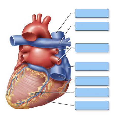
Correctly Label The Following External Anatomy Of The Posterior Heart.
Holbox
Apr 02, 2025 · 6 min read

Table of Contents
- Correctly Label The Following External Anatomy Of The Posterior Heart.
- Table of Contents
- Correctly Labeling the External Anatomy of the Posterior Heart
- Key Structures of the Posterior Heart: A Visual Guide
- 1. Left Atrium: The Dominant Feature
- 2. Right Atrium: A Smaller, Contributing Portion
- 3. Pulmonary Veins: The Oxygenated Blood Inlets
- 4. Superior and Inferior Vena Cava: Systemic Return
- 5. Esophagus and Azygos Vein: Adjacent Structures
- Precise Labeling Techniques: Mastering the Anatomy
- 1. Anatomical Dissection (Laboratory Setting)
- 2. High-Resolution Images and Illustrations: (Study)
- 3. Three-Dimensional Models: (Interactive learning)
- 4. Utilizing Cardiac Imaging Techniques (Clinical Setting)
- Common Errors to Avoid When Labeling
- Beyond Labeling: Understanding the Function
- Applying this Knowledge: Clinical Relevance
- Conclusion: Master the Posterior Heart
- Latest Posts
- Latest Posts
- Related Post
Correctly Labeling the External Anatomy of the Posterior Heart
The posterior aspect of the heart, often less discussed than the anterior surface, presents a unique anatomical landscape crucial for understanding cardiac function and pathology. Correctly identifying its features is fundamental for medical professionals, students, and anyone interested in cardiology. This comprehensive guide provides a detailed breakdown of the posterior heart's external anatomy, along with helpful tips for accurate labeling.
Key Structures of the Posterior Heart: A Visual Guide
Before delving into specifics, it's beneficial to visualize the posterior heart. Imagine the heart resting in the mediastinum, tilted slightly to the left. The posterior surface faces the vertebral column and is largely formed by the left atrium and parts of the right atrium. Several crucial structures define this surface:
1. Left Atrium: The Dominant Feature
The left atrium, a muscular chamber receiving oxygenated blood from the lungs via the pulmonary veins, dominates the posterior heart's anatomy. It's situated slightly posterior and superior to the other chambers. Its smooth posterior wall contrasts sharply with the rough, trabeculated walls of the atria’s interior. Understanding its size and shape is vital for assessing potential pathologies like atrial enlargement, often associated with heart failure.
- Clinical Significance: Enlargement of the left atrium is a significant indicator of various heart conditions, including mitral valve stenosis, mitral regurgitation, and hypertension.
2. Right Atrium: A Smaller, Contributing Portion
While less prominent than the left atrium on the posterior view, the right atrium does contribute to the posterior surface, particularly its superior portion. It's mainly involved in receiving deoxygenated blood from the systemic circulation through the superior and inferior vena cava. Observing the relative sizes and positions of the left and right atria can provide insights into overall cardiac health.
- Clinical Significance: Dilatation of the right atrium might suggest pulmonary hypertension or tricuspid valve dysfunction.
3. Pulmonary Veins: The Oxygenated Blood Inlets
Four pulmonary veins (two superior and two inferior) gracefully enter the left atrium, carrying the freshly oxygenated blood from the lungs. These veins are relatively short and often difficult to individually identify without careful dissection or imaging. However, their collective entrance point to the left atrium is clearly visible on the posterior heart.
- Clinical Significance: Abnormal pulmonary venous return can be a sign of various congenital heart defects or pulmonary diseases.
4. Superior and Inferior Vena Cava: Systemic Return
The superior and inferior vena cava are essential venous vessels that return deoxygenated blood from the systemic circulation to the right atrium. While primarily located on the anterior surface, their entrances into the right atrium are partially visible on the posterior view, particularly the inferior vena cava's opening near the atrioventricular junction.
- Clinical Significance: Blockages or abnormalities in the vena cava can lead to significant systemic venous congestion.
5. Esophagus and Azygos Vein: Adjacent Structures
While not directly part of the heart itself, the esophagus and the azygos vein lie in close proximity to the posterior heart, contributing to the anatomical landscape of the posterior mediastinum. The azygos vein, a significant vessel draining the posterior thoracic wall, is often visible during posterior heart examinations. The esophagus's position is crucial in considering potential complications from cardiac enlargement or surgical procedures.
- Clinical Significance: The relationship between these structures and the heart is relevant to surgical procedures, as well as to understanding symptoms related to compression or displacement.
Precise Labeling Techniques: Mastering the Anatomy
Accurately labeling the posterior heart requires a systematic approach:
1. Anatomical Dissection (Laboratory Setting)
For those with direct access to anatomical specimens, careful dissection under supervision is the most effective method. Using fine instruments and paying close attention to the delicate tissue around the pulmonary veins and great vessels is paramount. Each structure should be meticulously cleaned and examined before labeling.
2. High-Resolution Images and Illustrations: (Study)
Employing high-quality anatomical images and illustrations, either from textbooks or online resources, is crucial for studying the posterior heart's anatomy. These visual aids provide a detailed map, allowing for effective labeling practice. Look for images that clearly demonstrate the subtle relationships between different structures, like the angles of the pulmonary veins entering the left atrium.
3. Three-Dimensional Models: (Interactive learning)
Three-dimensional (3D) models offer a dynamic learning experience. By rotating and zooming in on the model, one can gain a comprehensive understanding of the heart's spatial organization and relationships between the various structures on the posterior surface. Interactive features on some 3D models can even simulate physiological processes, enhancing the learning experience.
4. Utilizing Cardiac Imaging Techniques (Clinical Setting)
In a clinical setting, various imaging techniques, including echocardiography and computed tomography (CT) scans, are used to visualize the posterior heart. Learning to interpret these images is crucial for medical professionals. Echo provides real-time visualization, enabling assessment of function alongside anatomy. CT scans offer detailed anatomical cross-sections, helpful for identifying complex relationships.
Common Errors to Avoid When Labeling
Several common pitfalls can lead to inaccurate labeling:
-
Confusing the Left and Right Atria: The left atrium's larger size and smooth posterior wall are key distinguishing features. Carefully noting these aspects prevents this common mistake.
-
Misidentifying Pulmonary Veins: The pulmonary veins' relatively small size and their entry points into the left atrium should be carefully observed. Their arrangement can sometimes be irregular, requiring detailed examination.
-
Ignoring Adjacent Structures: The relationships between the heart, esophagus, and azygos vein are essential to understanding the complete anatomical picture. Omitting these structures from the labeling process leads to an incomplete representation.
-
Insufficient Detail: Providing merely the names of the structures isn't sufficient. Include directional indicators (e.g., "Superior Vena Cava," "Inferior Pulmonary Vein, Left Side").
-
Lack of Context: Labeling should reflect the spatial arrangement of structures, reflecting their relative positions and connections to each other. This adds depth and demonstrates understanding.
Beyond Labeling: Understanding the Function
Correctly labeling the posterior heart’s anatomy is only the first step. Understanding the functional significance of each structure is critical. For instance, the left atrium's efficient contraction is essential for optimal blood flow to the systemic circulation. The precise arrangement of the pulmonary veins is crucial for efficient oxygenated blood delivery to the left atrium. A comprehensive understanding of both anatomy and physiology allows for a more holistic appreciation of cardiac function.
Applying this Knowledge: Clinical Relevance
The knowledge gained from accurately labeling the posterior heart is crucial for:
-
Diagnosing Cardiac Conditions: Identifying abnormalities in size, shape, or function of the posterior structures is key to diagnosing various heart conditions.
-
Cardiac Surgery Planning: Thorough knowledge of the posterior heart anatomy is crucial for planning and executing cardiovascular surgeries.
-
Interventional Cardiology: Accurate anatomical knowledge is paramount for procedures like catheterization and stent placement.
-
Medical Imaging Interpretation: The ability to correctly label structures in echocardiograms, CT scans, and other cardiac images is essential for accurate diagnosis and treatment planning.
Conclusion: Master the Posterior Heart
Mastering the external anatomy of the posterior heart is not merely an academic exercise; it's a foundation for understanding cardiac function, diagnosing disease, and performing various medical interventions. By using a combination of anatomical specimens, high-quality images, 3D models, and careful study techniques, one can develop a strong command of this crucial aspect of human anatomy. Remember to always focus on the precise relationships between structures and their functional significance. The more you practice, the more confident you will become in correctly labeling and understanding the posterior heart.
Latest Posts
Latest Posts
-
Label The Structures Of A Long Bone
Apr 06, 2025
-
What Are The Charges On Plates 3 And 6
Apr 06, 2025
-
What Product S Would You Expect From The Following Reaction
Apr 06, 2025
-
According To The Teachings Of The Buddha
Apr 06, 2025
-
You Need To Prepare An Acetate Buffer Of Ph
Apr 06, 2025
Related Post
Thank you for visiting our website which covers about Correctly Label The Following External Anatomy Of The Posterior Heart. . We hope the information provided has been useful to you. Feel free to contact us if you have any questions or need further assistance. See you next time and don't miss to bookmark.
