Correctly Label The Following Anatomical Parts Of A Kidney.
Holbox
Apr 02, 2025 · 8 min read
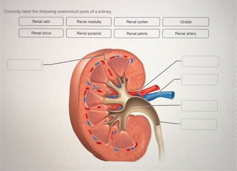
Table of Contents
- Correctly Label The Following Anatomical Parts Of A Kidney.
- Table of Contents
- Correctly Labeling the Anatomical Parts of a Kidney: A Comprehensive Guide
- External Anatomy of the Kidney: The Macro View
- 1. Renal Capsule: The Protective Outer Layer
- 2. Renal Hilum: The Gateway to the Kidney
- 3. Renal Fascia: Anchoring the Kidney
- 4. Perirenal Fat Capsule: Cushioning and Protection
- 5. Adipose Capsule: Additional Protection
- Internal Anatomy of the Kidney: A Microscopic Journey
- 1. Renal Cortex: The Outer Region
- 2. Renal Medulla: The Inner Region
- 3. Renal Pyramids: Triangular Structures
- 4. Renal Papilla: Urine Collection Point
- 5. Minor Calyces: Initial Urine Collection
- 6. Major Calyces: Consolidation of Urine
- 7. Renal Pelvis: The Urine Reservoir
- 8. Ureter: Transporting Urine
- Nephron: The Functional Unit of the Kidney
- 1. Renal Corpuscle: The Filtration Site
- 2. Renal Tubule: Processing the Filtrate
- Blood Supply to the Kidney: A Complex Network
- 1. Renal Artery: The Main Supply Line
- 2. Segmental Arteries: Distributing Blood
- 3. Interlobar Arteries: Towards the Cortex
- 4. Arcuate Arteries: Arching Across the Pyramids
- 5. Interlobular Arteries: Blood to the Nephrons
- 6. Glomerular Capillaries: Filtration Site
- 7. Efferent Arterioles: Blood Flow Regulation
- 8. Peritubular Capillaries: Reabsorption and Secretion
- 9. Renal Vein: Blood Exit Route
- Clinical Significance and Importance
- Latest Posts
- Latest Posts
- Related Post
Correctly Labeling the Anatomical Parts of a Kidney: A Comprehensive Guide
The kidney, a vital organ in the urinary system, plays a crucial role in maintaining overall health and homeostasis. Understanding its intricate anatomy is essential for anyone studying biology, medicine, or related fields. This comprehensive guide will delve deep into the detailed structure of the kidney, providing a clear and concise explanation of each component, along with high-quality imagery (which cannot be displayed here but is strongly recommended to use alongside this guide for best understanding). This guide aims to equip you with the knowledge to correctly label all major anatomical parts of a kidney.
External Anatomy of the Kidney: The Macro View
Before we delve into the microscopic details, let's begin by examining the kidney's external features. A typical human kidney, roughly the size and shape of a fist, exhibits several key characteristics:
1. Renal Capsule: The Protective Outer Layer
The renal capsule is a tough, fibrous membrane that directly encases the kidney. It provides vital protection against physical trauma and infection, acting as the first line of defense for this delicate organ. Its smooth, glistening surface contributes to the kidney's overall appearance.
2. Renal Hilum: The Gateway to the Kidney
Located on the medial concave border of the kidney, the renal hilum is a significant indentation that serves as the entry and exit point for several crucial structures. This includes the renal artery, renal vein, and ureter.
3. Renal Fascia: Anchoring the Kidney
Surrounding the kidney externally is the renal fascia, a layer of connective tissue that anchors the kidney securely in place within the abdominal cavity. This fascia helps to maintain the kidney's proper position and prevent excessive movement.
4. Perirenal Fat Capsule: Cushioning and Protection
Between the renal fascia and the renal capsule lies the perirenal fat capsule. This layer of adipose tissue provides excellent cushioning and insulation, protecting the kidney from physical shocks and temperature fluctuations. Its thickness varies considerably depending on an individual's overall body fat percentage.
5. Adipose Capsule: Additional Protection
External to the renal fascia lies the adipose capsule, a layer of fat that provides additional cushioning and support to the kidney within the abdominal cavity. This layer helps to maintain the kidney's position and protect it against trauma.
Internal Anatomy of the Kidney: A Microscopic Journey
Now, let's move inward to explore the kidney's internal structure, which is equally complex and fascinating. This area is where the vital process of filtration and urine formation takes place.
1. Renal Cortex: The Outer Region
The renal cortex, the outermost layer of the kidney's internal structure, has a granular appearance due to the presence of numerous nephrons, the functional units of the kidney. It's here that the initial stages of blood filtration occur. The cortex extends inward as renal columns between the pyramids.
2. Renal Medulla: The Inner Region
Deep within the cortex lies the renal medulla, a darker, striated region composed of cone-shaped structures called renal pyramids. The pyramids are organized in a manner that creates distinct renal columns between them. The medulla plays a crucial role in concentrating urine.
3. Renal Pyramids: Triangular Structures
The renal pyramids are triangular-shaped structures within the medulla. These structures contain the loops of Henle and collecting ducts of nephrons, key components in the urine concentration process. The apex of each pyramid points towards the renal papilla.
4. Renal Papilla: Urine Collection Point
The renal papilla is the apex of each renal pyramid. It projects into a minor calyx, the initial collecting point for the urine formed within the pyramid. This structure marks the transition from the medullary area to the collecting system.
5. Minor Calyces: Initial Urine Collection
Several minor calyces (singular: calyx) converge to form a larger structure. These cup-like structures collect urine from the renal papillae. This is where urine begins its journey toward the ureter.
6. Major Calyces: Consolidation of Urine
The major calyces are larger structures formed by the fusion of multiple minor calyces. They further consolidate the urine before delivering it to the renal pelvis.
7. Renal Pelvis: The Urine Reservoir
The renal pelvis is a funnel-shaped structure that collects urine from the major calyces. It acts as a reservoir before the urine is passed into the ureter for transport to the bladder.
8. Ureter: Transporting Urine
The ureter is a long, muscular tube that extends from the renal pelvis to the urinary bladder. Its rhythmic contractions, called peristalsis, propel the urine towards the bladder for storage and eventual elimination from the body.
Nephron: The Functional Unit of the Kidney
The nephron is the microscopic functional unit of the kidney, responsible for filtering blood and producing urine. Millions of nephrons are packed within each kidney, allowing for efficient processing of vast amounts of blood. Understanding the nephron's structure is crucial for grasping the kidney's overall function.
1. Renal Corpuscle: The Filtration Site
The renal corpuscle is the initial filtration unit of the nephron. It consists of two main structures:
- Glomerulus: A network of capillaries where blood filtration takes place.
- Bowman's Capsule: A double-walled cup-like structure that surrounds the glomerulus and collects the filtrate.
2. Renal Tubule: Processing the Filtrate
The renal tubule is a long, convoluted tube that processes the filtrate from Bowman's capsule. It's divided into several segments:
- Proximal Convoluted Tubule (PCT): The first segment, reabsorbing essential nutrients and water.
- Loop of Henle: A U-shaped structure extending into the medulla, playing a critical role in concentrating urine.
- Distal Convoluted Tubule (DCT): The final segment, involved in fine-tuning electrolyte balance.
- Collecting Duct: A shared duct that receives filtrate from multiple nephrons, further concentrating the urine before it reaches the renal papilla.
Blood Supply to the Kidney: A Complex Network
The kidney's intricate function necessitates a robust and efficient blood supply. Understanding the vascular network is essential for comprehending how blood is filtered and processed within the nephrons.
1. Renal Artery: The Main Supply Line
The renal artery, a branch of the abdominal aorta, delivers oxygenated blood to the kidney. It enters the kidney through the renal hilum and branches extensively to supply all parts of the organ.
2. Segmental Arteries: Distributing Blood
The renal artery branches into several segmental arteries, distributing blood to different segments of the kidney. These arteries further divide into smaller vessels to ensure adequate perfusion of all nephrons.
3. Interlobar Arteries: Towards the Cortex
The segmental arteries then branch into interlobar arteries, traveling between the renal pyramids. These arteries play a crucial role in distributing blood to the renal cortex.
4. Arcuate Arteries: Arching Across the Pyramids
At the boundary between the cortex and medulla, the interlobar arteries give rise to arcuate arteries, which arch over the renal pyramids.
5. Interlobular Arteries: Blood to the Nephrons
The arcuate arteries branch into interlobular arteries, supplying blood to the nephrons themselves. These vessels form the afferent arterioles that lead to the glomeruli.
6. Glomerular Capillaries: Filtration Site
The glomerular capillaries, part of the renal corpuscle, are where blood filtration occurs. They are highly permeable and allow for the passage of water and small solutes into Bowman's capsule.
7. Efferent Arterioles: Blood Flow Regulation
The efferent arterioles carry blood away from the glomerulus. Their diameter influences glomerular filtration rate (GFR).
8. Peritubular Capillaries: Reabsorption and Secretion
The peritubular capillaries surround the renal tubules and are involved in reabsorption of essential substances and secretion of waste products.
9. Renal Vein: Blood Exit Route
The renal vein carries deoxygenated blood from the kidney back to the inferior vena cava, completing the circulatory loop. It leaves the kidney via the renal hilum alongside the renal artery and ureter.
Clinical Significance and Importance
A thorough understanding of the kidney's anatomy is essential in various clinical settings. Accurate labeling of its parts is crucial for:
-
Diagnosing and treating kidney diseases: Identifying abnormalities in the kidney's structure can pinpoint the cause of various renal disorders. Imaging techniques, such as ultrasounds, CT scans, and MRIs, rely on a strong anatomical knowledge for accurate interpretation.
-
Performing kidney surgeries: Surgeons require detailed knowledge of the kidney's intricate structure for successful procedures like nephrectomies (kidney removal) or partial nephrectomies.
-
Understanding the effects of medications: Many medications are metabolized and excreted by the kidneys, making anatomical understanding crucial for prescribing and monitoring drug therapies.
-
Evaluating kidney function tests: Analyzing blood and urine samples requires knowledge of the kidney's anatomical structures and their functions to accurately interpret the results.
In conclusion, accurately labeling the anatomical parts of a kidney requires a comprehensive understanding of both its external and internal structures, as well as its complex vascular network and the functional units (nephrons) responsible for urine production. This detailed knowledge forms a solid foundation for advancing medical understanding and improving patient care. Remember to utilize anatomical diagrams and models for visual learning and reinforcement. The detail presented here provides a robust groundwork for correctly labeling the various components of this vital organ.
Latest Posts
Latest Posts
-
Which Empty Cleaned And Sanitized Container
Apr 05, 2025
-
Decision Making Management Information Systems Are Necessary Because
Apr 05, 2025
-
Which Of The Following Statements About The Cytoskeleton Is False
Apr 05, 2025
-
Identify The Relationship Between The Following Compounds
Apr 05, 2025
-
What Proportion Of Employees Have A Pc
Apr 05, 2025
Related Post
Thank you for visiting our website which covers about Correctly Label The Following Anatomical Parts Of A Kidney. . We hope the information provided has been useful to you. Feel free to contact us if you have any questions or need further assistance. See you next time and don't miss to bookmark.
