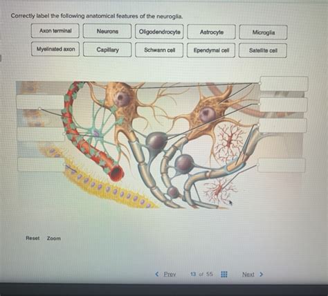
Correctly Label The Following Anatomical Features Of The Neuroglia
Holbox
Mar 31, 2025 · 6 min read

Table of Contents
- Correctly Label The Following Anatomical Features Of The Neuroglia
- Table of Contents
- Correctly Labeling the Anatomical Features of Neuroglia: A Comprehensive Guide
- Major Types of Neuroglia and Their Defining Characteristics
- 1. Astrocytes (CNS)
- 2. Oligodendrocytes (CNS)
- 3. Microglia (CNS)
- 4. Ependymal Cells (CNS)
- 5. Schwann Cells (PNS)
- 6. Satellite Cells (PNS)
- Practical Applications of Accurate Neuroglia Labeling
- Advanced Techniques in Neuroglia Labeling
- Conclusion
- Latest Posts
- Latest Posts
- Related Post
Correctly Labeling the Anatomical Features of Neuroglia: A Comprehensive Guide
Neuroglia, also known as glial cells, are non-neuronal cells in the central nervous system (CNS) and the peripheral nervous system (PNS). They provide support and protection for neurons, contributing significantly to the overall function and health of the nervous system. While neurons are the stars of the show, responsible for transmitting nerve impulses, neuroglia are the essential supporting cast, ensuring the proper functioning of the entire system. This article will delve into the detailed anatomy of various neuroglia, focusing on accurate labeling of their key features. Understanding these structures is crucial for comprehending the complex interactions within the nervous system.
Major Types of Neuroglia and Their Defining Characteristics
Before diving into specific anatomical features, it's crucial to understand the different types of neuroglia. They are broadly classified into several categories based on their location and function:
1. Astrocytes (CNS)
Astrocytes, meaning "star cells," are the most abundant glial cells in the CNS. Their star-shaped morphology is immediately recognizable. Their key functions include:
- Structural Support: They provide a physical framework for neurons, holding them in place.
- Blood-Brain Barrier (BBB) Regulation: Astrocyte foot processes are crucial in forming and maintaining the BBB, a selective barrier that protects the brain from harmful substances in the blood. Labeling this would involve identifying the end-feet of astrocytes in close proximity to blood vessels.
- Neurotransmitter Recycling: They help regulate the synaptic cleft by removing excess neurotransmitters, thus ensuring efficient neuronal signaling. This would require labeling the processes that extend to synapses.
- Nutrient Supply: They provide metabolic support to neurons by delivering nutrients and removing waste products. This function would be represented by labeling the processes that connect to neurons and blood vessels.
- Synaptic Plasticity: Recent research highlights their role in synaptic plasticity, influencing the strength and efficiency of neuronal connections.
Anatomical Features to Label:
- Cell Body (Soma): The central part of the astrocyte containing the nucleus and organelles.
- Processes: Numerous long, branching processes extending from the soma.
- End-feet: Specialized processes that contact blood vessels and form part of the BBB.
- Gap Junctions: Connections between astrocytes that allow for communication and coordination.
2. Oligodendrocytes (CNS)
Oligodendrocytes are responsible for myelination in the CNS. Myelin is a fatty insulating sheath that surrounds axons, increasing the speed of nerve impulse conduction.
Anatomical Features to Label:
- Cell Body: Relatively small and compact compared to astrocytes.
- Myelin Sheath: The characteristic fatty layers formed by oligodendrocytes wrapping around axons. Labeling should clearly indicate the concentric layers of the myelin.
- Internodes: The segments of the axon covered by myelin.
- Nodes of Ranvier: The gaps between myelin sheaths where the axon is exposed. These are crucial for saltatory conduction.
- Multiple Axonal Segments: A single oligodendrocyte can myelinate multiple segments of different axons. This is a key differentiator from Schwann cells.
3. Microglia (CNS)
Microglia are the resident immune cells of the CNS. They act as the brain's first line of defense against infection and injury.
Anatomical Features to Label:
- Small Cell Body: They have a small, elongated cell body.
- Highly Branched Processes: Their processes are highly dynamic and ramified, constantly surveying their environment.
- Amoeboid Movement: During immune responses, microglia can transform into an amoeboid form to engulf pathogens and cellular debris. Labeling might focus on the characteristic changes in cell shape.
- Phagocytic Vesicles: These vesicles are visible when microglia are actively engulfing debris.
4. Ependymal Cells (CNS)
Ependymal cells line the ventricles of the brain and the central canal of the spinal cord. They are involved in the production and circulation of cerebrospinal fluid (CSF).
Anatomical Features to Label:
- Cuboidal or Columnar Shape: They have a characteristic epithelial-like structure.
- Cilia: Many ependymal cells possess cilia, which help to circulate CSF. Labeling should clearly show the hair-like projections.
- Microvilli: Some ependymal cells have microvilli, which may be involved in absorption or secretion.
- Tight Junctions: These junctions between ependymal cells help to form the blood-CSF barrier.
5. Schwann Cells (PNS)
Schwann cells are the myelinating cells of the peripheral nervous system (PNS). Similar to oligodendrocytes, they produce myelin sheaths around axons, but with some key differences.
Anatomical Features to Label:
- Cell Body (Soma): Contains the nucleus and organelles.
- Myelin Sheath: Forms the myelin sheath around a single axon segment. This is a significant difference from oligodendrocytes.
- Neurilemma: The outer layer of the Schwann cell, which wraps around the myelin sheath. It is also called the Schwann cell sheath.
- Nodes of Ranvier: Similar to those in the CNS, these gaps between myelin sheaths are crucial for fast nerve impulse conduction.
- Single Axonal Segment: Each Schwann cell myelinated only one segment of a single axon.
6. Satellite Cells (PNS)
Satellite cells surround neuron cell bodies in ganglia of the PNS. They provide structural support and regulate the microenvironment around neurons.
Anatomical Features to Label:
- Flattened Cell Body: They are small and flattened cells surrounding neuronal cell bodies.
- Processes: These processes extend between the neurons they surround.
- Close Association with Neurons: Their close proximity to neuron cell bodies indicates their crucial support role.
Practical Applications of Accurate Neuroglia Labeling
Accurate labeling of neuroglia is essential in various contexts:
- Neuroscience Research: Understanding the precise structure and function of glial cells is fundamental to advancing neuroscience research. Accurate labeling is crucial in studies investigating neurological disorders, brain development, and neuronal plasticity.
- Medical Diagnosis: Microscopic analysis of brain tissue relies on the ability to identify different types of neuroglia. Misidentification can lead to inaccurate diagnoses and treatment plans. Neuroglia often exhibit characteristic changes in disease states, such as inflammation or demyelination, and the specific changes must be correctly identified.
- Drug Development: Many neurodegenerative diseases and CNS disorders affect neuroglia. Therefore, accurate identification of neuroglia is critical in the development of new therapeutic agents that target specific glial cell functions.
- Educational Purposes: Accurate labeling and illustration of neuroglia are essential for teaching and understanding the structure and function of the nervous system. Clear visualization helps students understand the complexities of the CNS and PNS.
Advanced Techniques in Neuroglia Labeling
Several advanced techniques are used to visualize and label neuroglia for research and diagnostic purposes:
- Immunohistochemistry: This technique uses specific antibodies to label particular proteins found in different types of neuroglia. This allows for precise identification of cell types.
- In Situ Hybridization: This method uses labeled probes to detect specific mRNA sequences, indicating the expression of particular genes within neuroglia.
- Confocal Microscopy: This technique provides high-resolution 3D images of neuroglia, allowing for a detailed examination of their morphology and interactions with other cells.
- Electron Microscopy: This provides extremely high-resolution images, revealing the fine details of neuroglial structure, such as myelin sheaths and cell junctions.
Conclusion
Correctly labeling the anatomical features of neuroglia is crucial for understanding the complex workings of the nervous system. The detailed structural characteristics discussed above, coupled with advanced imaging techniques, provide a powerful toolbox for neuroscientists, clinicians, and educators alike. As our understanding of neuroglia continues to evolve, so will the methods employed to accurately visualize and classify these vital cells. The detailed knowledge of neuroglial anatomy is foundational to advancements in neuroscience research, medical diagnostics, and the development of effective treatments for neurological disorders.
Latest Posts
Latest Posts
-
What Defines A Long Term Investment Check All That Apply
Apr 04, 2025
-
A Market Product Grid Is A Framework To Relate
Apr 04, 2025
-
The X Coordinate Of An Ordered Pair Specifies The
Apr 04, 2025
-
Organisms That Extract Energy From Nonliving Environmental Resources Are Called
Apr 04, 2025
-
The Body Of A Direct Request Letter Should
Apr 04, 2025
Related Post
Thank you for visiting our website which covers about Correctly Label The Following Anatomical Features Of The Neuroglia . We hope the information provided has been useful to you. Feel free to contact us if you have any questions or need further assistance. See you next time and don't miss to bookmark.
