Correctly Label The Following Anatomical Features Of A Nerve
Holbox
Mar 24, 2025 · 6 min read
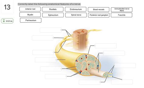
Table of Contents
- Correctly Label The Following Anatomical Features Of A Nerve
- Table of Contents
- Correctly Labeling the Anatomical Features of a Nerve: A Comprehensive Guide
- The Basic Structure of a Nerve
- 1. Endoneurium: The Innermost Layer
- 2. Perineurium: Bundling the Axons
- 3. Epineurium: The Outermost Sheath
- Key Anatomical Features to Label
- 1. Axons: The Signal Transmitters
- 2. Myelin Sheath: The Insulating Layer
- 3. Nodes of Ranvier: Facilitating Fast Conduction
- 4. Schwann Cells/Oligodendrocytes: Myelinating Cells
- 5. Neurolemma: The Outermost Layer of Schwann Cells
- 6. Fascicles: Bundles of Axons
- 7. Perineurium: The Fascicle's Protective Sheath
- 8. Epineurium: The Outermost Covering
- 9. Blood Vessels: Providing Nutrients and Oxygen
- 10. Connective Tissue: Providing Structural Support
- 11. Sensory and Motor Nerve Fibers: Functional Distinction
- 12. Myelinated and Unmyelinated Axons: Conduction Speed Differences
- 13. Nerve Branches/Collateral Branches: Diversification of Signals
- Practical Applications of Accurate Nerve Labeling
- Conclusion: The Importance of Precision
- Latest Posts
- Latest Posts
- Related Post
Correctly Labeling the Anatomical Features of a Nerve: A Comprehensive Guide
Understanding the intricate anatomy of a nerve is crucial for anyone studying neurobiology, medicine, or related fields. Nerves, the communication highways of the body, are complex structures with various components that work together to transmit signals. Correctly identifying and labeling these features is essential for accurate diagnosis, treatment, and research. This comprehensive guide will delve into the key anatomical features of a nerve, providing detailed descriptions and aiding in their accurate labeling.
The Basic Structure of a Nerve
Before diving into specific features, let's establish a foundational understanding of nerve structure. A nerve is essentially a bundle of nerve fibers, also known as axons, held together by various connective tissues. These axons are the long projections of neurons, specialized cells that transmit electrical and chemical signals throughout the body. The axons are not solitary; they are organized and protected by several layers of connective tissue:
1. Endoneurium: The Innermost Layer
The endoneurium is a delicate layer of connective tissue that surrounds each individual axon. It's composed of collagen fibers, fibroblasts, and a basement membrane, providing structural support and insulation to each axon. Think of it as the individual insulation around each wire in a cable.
2. Perineurium: Bundling the Axons
Multiple axons grouped together form a fascicle. The perineurium is a thicker, more robust layer of connective tissue that encases each fascicle. This layer is crucial for protecting the axons within and creating a barrier against the spread of infection or inflammation. The perineurium is composed of concentric layers of flattened cells, creating a strong, yet flexible structure.
3. Epineurium: The Outermost Sheath
The epineurium is the outermost layer of connective tissue, enveloping the entire nerve. It's the thickest and most substantial layer, providing the overall structure and protection for the nerve. It contains blood vessels and lymphatic channels that supply nutrients and remove waste products from the nerve fibers. The epineurium also plays a significant role in maintaining the nerve's shape and preventing damage from external forces.
Key Anatomical Features to Label
Now, let's delve into specific anatomical features that often require labeling in anatomical diagrams and studies. Accurate labeling is essential for clear communication and understanding.
1. Axons: The Signal Transmitters
Axons are the fundamental units of nerve transmission. They are long, slender projections of neurons responsible for carrying nerve impulses away from the cell body (soma). Labeling an axon should clearly distinguish it from other components, highlighting its cylindrical shape and its role in signal propagation. Different types of axons may vary in diameter and myelination, features also worth noting if appropriate.
2. Myelin Sheath: The Insulating Layer
Many axons are surrounded by a myelin sheath, a fatty insulating layer that greatly increases the speed of nerve impulse conduction. This sheath is formed by specialized glial cells: oligodendrocytes in the central nervous system (CNS) and Schwann cells in the peripheral nervous system (PNS). The myelin sheath is not continuous; it's segmented, with gaps called Nodes of Ranvier between the segments. These nodes play a crucial role in saltatory conduction, a rapid form of signal transmission.
3. Nodes of Ranvier: Facilitating Fast Conduction
Nodes of Ranvier are the periodic gaps in the myelin sheath. Their presence is essential for saltatory conduction, a process where the nerve impulse "jumps" from node to node, significantly increasing the speed of signal transmission. In diagrams, these should be clearly indicated as gaps in the myelin sheath.
4. Schwann Cells/Oligodendrocytes: Myelinating Cells
Clearly labeling the cells responsible for myelin production is crucial. In the PNS, these are Schwann cells, while in the CNS, they are oligodendrocytes. Distinguishing between the two is important because they differ in their morphology and function. A detailed label should specify which type of glial cell is present.
5. Neurolemma: The Outermost Layer of Schwann Cells
The neurolemma, also known as the Schwann cell sheath, is the outermost layer of the Schwann cell surrounding the axon. It remains intact even after myelin degradation, playing a role in axon regeneration. This feature is especially important in understanding nerve repair mechanisms.
6. Fascicles: Bundles of Axons
Fascicles are bundles of axons grouped together within the nerve. Their boundaries are clearly defined by the perineurium. When labeling a fascicle, it's useful to indicate the presence of the perineurium surrounding it.
7. Perineurium: The Fascicle's Protective Sheath
The perineurium is the connective tissue layer that surrounds each fascicle. This layer provides structural support and a barrier to protect the axons within. The labeling should highlight its layered structure and its role in nerve protection.
8. Epineurium: The Outermost Covering
The epineurium is the outermost layer of connective tissue, encompassing the entire nerve. It provides the overall structure and protection for the nerve, containing blood vessels and lymphatic channels. Labeling this layer should emphasize its role in providing vascular support and overall nerve protection.
9. Blood Vessels: Providing Nutrients and Oxygen
Nerves require a constant supply of oxygen and nutrients to function properly. Blood vessels within the epineurium and perineurium deliver these vital substances. Labeling these vessels is essential for understanding nerve physiology and metabolism.
10. Connective Tissue: Providing Structural Support
The various layers of connective tissue (endoneurium, perineurium, and epineurium) provide essential structural support to the nerve. Highlighting their different roles in maintaining nerve integrity is important for a complete anatomical description. Mentioning the types of connective tissue fibers (e.g., collagen) can enhance the label's accuracy.
11. Sensory and Motor Nerve Fibers: Functional Distinction
Nerves often contain a mixture of sensory and motor nerve fibers. Sensory fibers transmit information from the periphery to the CNS, while motor fibers transmit signals from the CNS to muscles or glands. Distinguishing between these fiber types in a label provides crucial functional information.
12. Myelinated and Unmyelinated Axons: Conduction Speed Differences
Axons can be either myelinated or unmyelinated. Myelinated axons conduct signals faster due to saltatory conduction. Clearly indicating the presence of both types within a nerve provides valuable physiological context.
13. Nerve Branches/Collateral Branches: Diversification of Signals
Nerves often branch to reach their target tissues. These branches, also called collateral branches, allow the nerve to innervate multiple areas or structures. Labeling these branches helps visualize the nerve's distribution and function.
Practical Applications of Accurate Nerve Labeling
Accurate labeling of nerve anatomical features is not merely an academic exercise; it has significant practical applications in various fields:
- Neurosurgery: Precise identification of nerve components is crucial during surgical procedures to minimize damage and ensure optimal outcomes.
- Neurological Diagnosis: Accurate labeling assists in the diagnosis of neurological disorders by allowing for a clear understanding of the affected structures.
- Neuropathology: Detailed labeling helps in analyzing nerve tissue samples and identifying pathological changes associated with diseases.
- Neurological Research: Accurate labeling is essential for conducting research on nerve function, regeneration, and repair.
- Medical Illustration and Education: Clear and accurate anatomical diagrams with correct labels are invaluable for educating medical students and other healthcare professionals.
Conclusion: The Importance of Precision
Correctly labeling the anatomical features of a nerve is paramount for a thorough understanding of its structure and function. This guide provided a detailed overview of the key components, emphasizing the importance of precision in labeling. Mastering this skill is essential for anyone working in fields related to neurobiology, medicine, or neuroscience research. The ability to accurately identify and label nerve structures not only enhances comprehension but also contributes to the advancement of diagnostic and therapeutic techniques in the field of neurology. By understanding the intricate details of nerve anatomy, we can better appreciate the complexity and critical role of the nervous system in maintaining overall health and function.
Latest Posts
Latest Posts
-
Label The Structures Of The Urinary Tract In The Figure
Mar 26, 2025
-
Which Of The Following Is A Function Of Protein
Mar 26, 2025
-
Campaigning Its A Process Answer Key
Mar 26, 2025
-
Rn Pharmacology Online Practice 2023 B
Mar 26, 2025
-
A Sales Rep Is Displaying His Companys Newest Smartwatches
Mar 26, 2025
Related Post
Thank you for visiting our website which covers about Correctly Label The Following Anatomical Features Of A Nerve . We hope the information provided has been useful to you. Feel free to contact us if you have any questions or need further assistance. See you next time and don't miss to bookmark.
