Correctly Label The Anterior Muscles Of The Thigh
Holbox
Mar 18, 2025 · 5 min read
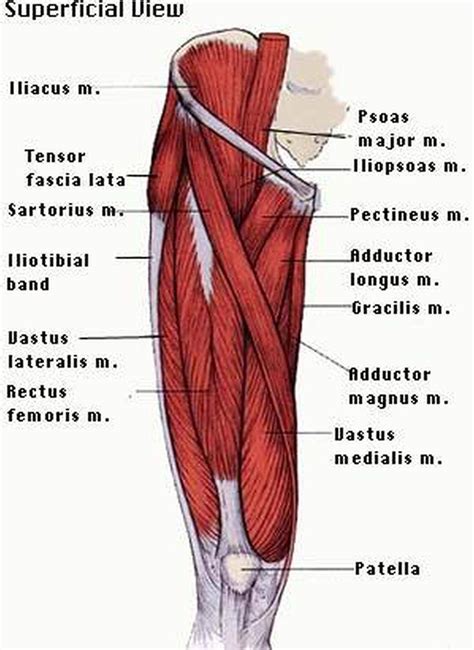
Table of Contents
Correctly Labeling the Anterior Muscles of the Thigh: A Comprehensive Guide
The anterior compartment of the thigh houses a group of muscles crucial for hip flexion, knee extension, and various other movements essential for daily activities and athletic performance. Accurate identification of these muscles is vital for healthcare professionals, fitness instructors, and anyone interested in human anatomy. This comprehensive guide will delve into the specifics of each muscle, providing detailed descriptions, origins and insertions, actions, and clinically relevant information. We will also discuss common mistakes in identification and provide tips for accurate labeling.
Understanding the Anterior Compartment
Before we delve into the individual muscles, let's establish the context. The anterior compartment of the thigh is located at the front of the thigh, bordered by the inguinal ligament superiorly, the patella inferiorly, and the medial and lateral intermuscular septa laterally and medially. This compartment is primarily responsible for extending the knee and flexing the hip. Its key players include the quadriceps femoris muscle group and the sartorius muscle.
The Quadriceps Femoris: The Powerhouse of Knee Extension
The quadriceps femoris is not a single muscle but a group of four muscles – rectus femoris, vastus lateralis, vastus medialis, and vastus intermedius – that converge to form the quadriceps tendon, which inserts into the tibial tuberosity via the patella. Let's explore each individually:
Rectus Femoris: The Two-Joint Muscle
- Origin: Anterior inferior iliac spine (AIIS) and superior acetabulum. Note the unique dual origin, making it the only quadriceps muscle to cross both the hip and knee joints.
- Insertion: Tibial tuberosity via the quadriceps and patellar tendons.
- Action: Flexes the hip and extends the knee. Its hip flexion action is particularly important during activities like kicking a ball or lifting the leg.
Clinical Relevance: Strains to the rectus femoris are common, particularly in athletes involved in sprinting or kicking sports. These strains can manifest as pain in the anterior thigh and groin region.
Vastus Lateralis: The Largest of the Quadriceps
- Origin: Greater trochanter, intertrochanteric line, linea aspera, and lateral supracondylar ridge of the femur.
- Insertion: Tibial tuberosity via the quadriceps and patellar tendons.
- Action: Extends the knee. Its powerful action is crucial for activities such as walking, running, and jumping.
Clinical Relevance: The vastus lateralis is a common site for intramuscular injections. Pain or weakness in this muscle can be indicative of various conditions, including muscle strains, tendinitis, and referred pain from other sources.
Vastus Medialis: Medial Knee Stabilizer
- Origin: Intertrochanteric line, medial linea aspera, and medial supracondylar line of the femur.
- Insertion: Tibial tuberosity via the quadriceps and patellar tendons.
- Action: Extends the knee. Plays a crucial role in stabilizing the patella and maintaining proper knee tracking.
Clinical Relevance: Weakness or dysfunction of the vastus medialis obliquus (VMO), a specific part of the vastus medialis, is often implicated in patellofemoral pain syndrome (runner's knee).
Vastus Intermedius: Deepest of the Quadriceps
- Origin: Anterior and lateral surfaces of the femur.
- Insertion: Tibial tuberosity via the quadriceps and patellar tendons.
- Action: Extends the knee. Often difficult to palpate due to its deep position, making accurate labeling a challenge.
Clinical Relevance: Because of its deep location, injuries to the vastus intermedius are often less apparent than injuries to the other quadriceps muscles.
Sartorius: The Tailor's Muscle
The sartorius muscle, despite being located in the anterior compartment, is unique due to its long, strap-like structure and its action across multiple joints.
- Origin: Anterior superior iliac spine (ASIS).
- Insertion: Medial surface of the proximal tibia (pes anserinus).
- Action: Flexes, abducts, and laterally rotates the hip; flexes the knee. Its actions contribute to a variety of movements, including crossing the legs (hence the "tailor's muscle" moniker).
Clinical Relevance: The sartorius is sometimes involved in strains or tears, particularly in activities involving rapid changes in direction or forceful hip movements.
Common Mistakes in Labeling Anterior Thigh Muscles
Accurate labeling requires meticulous observation and understanding of anatomical landmarks. Here are some common errors:
- Confusing Vastus Medialis and Vastus Lateralis: The vastus lateralis is significantly larger and more easily palpable than the vastus medialis. Careful palpation and anatomical knowledge are crucial to differentiate between them.
- Misidentifying Vastus Intermedius: The deep location of the vastus intermedius makes it challenging to identify visually or by palpation. Knowledge of its origin and insertion points is essential.
- Overlooking the Rectus Femoris' Dual Origin: Failure to recognize the rectus femoris' origins at both the AIIS and superior acetabulum is a frequent error.
- Incorrectly Assigning Actions: Understanding the actions of each muscle—specifically, the rectus femoris' role in both hip flexion and knee extension—is crucial for accurate labeling.
Tips for Accurate Labeling
To ensure correct identification, consider the following:
- Systematic Approach: Begin by identifying easily palpable landmarks like the ASIS, greater trochanter, and patella.
- Palpation: Carefully palpate each muscle to appreciate its shape, size, and location.
- Anatomical Charts: Refer to detailed anatomical charts and diagrams to visualize the muscle origins, insertions, and relative positions.
- Clinical Correlation: Consider potential clinical presentations associated with each muscle to aid in identification.
- Comparative Anatomy: Compare the muscle's features to those of neighbouring muscles to distinguish them.
Conclusion
Correctly labeling the anterior muscles of the thigh requires a thorough understanding of their anatomical characteristics and functional roles. By following the detailed descriptions and tips provided in this guide and integrating multiple learning methods such as palpation and visual aids, you can confidently identify the rectus femoris, vastus lateralis, vastus medialis, vastus intermedius, and sartorius muscles. This comprehensive knowledge is invaluable for healthcare professionals, fitness enthusiasts, and anyone interested in the intricacies of human anatomy. Remember to practice regularly and consult reliable anatomical resources to solidify your understanding and ensure accurate labeling. Mastering this skill opens doors to deeper comprehension of movement, injury, and rehabilitation strategies.
Latest Posts
Latest Posts
-
The Most Common Output Device For Soft Output Is A
Mar 18, 2025
-
A Clarinetist Setting Out For A Performance
Mar 18, 2025
-
645 Is The Same As What Percent
Mar 18, 2025
-
How To Cite Survey In Mla
Mar 18, 2025
-
Specific Performance Is A Remedy Which Is Always Available In
Mar 18, 2025
Related Post
Thank you for visiting our website which covers about Correctly Label The Anterior Muscles Of The Thigh . We hope the information provided has been useful to you. Feel free to contact us if you have any questions or need further assistance. See you next time and don't miss to bookmark.
