Correctly Label The Anatomical Features Of A Neuromuscular Junction
Holbox
Apr 02, 2025 · 6 min read
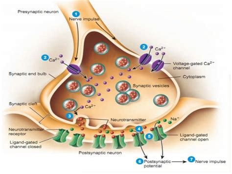
Table of Contents
- Correctly Label The Anatomical Features Of A Neuromuscular Junction
- Table of Contents
- Correctly Labeling the Anatomical Features of a Neuromuscular Junction
- Key Players: The Motor Neuron and the Muscle Fiber
- 1. The Motor Neuron: The Messenger
- 2. The Muscle Fiber: The Responder
- The Neuromuscular Junction: A Detailed Anatomy
- 1. The Axon Terminal: The Transmitter Release Zone
- 2. The Synaptic Cleft: The Communication Bridge
- 3. The Motor End Plate: The Receptor-Rich Zone
- 4. Schwann Cells: The Protective Sheath
- 5. Basal Lamina: The Supporting Structure
- Labeling the NMJ: A Step-by-Step Guide
- Clinical Significance of Understanding the NMJ
- Conclusion: Mastering the NMJ Anatomy
- Latest Posts
- Latest Posts
- Related Post
Correctly Labeling the Anatomical Features of a Neuromuscular Junction
The neuromuscular junction (NMJ), also known as the myoneural junction, is a highly specialized chemical synapse between a motor neuron and a muscle fiber. It's the site where the nervous system communicates with muscles, triggering muscle contraction. Understanding its intricate anatomy is crucial for grasping the mechanisms of movement, and for diagnosing and treating neuromuscular diseases. This article provides a comprehensive guide to correctly labeling the key anatomical features of the neuromuscular junction.
Key Players: The Motor Neuron and the Muscle Fiber
Before diving into the specific components of the NMJ, it's essential to establish the two major players:
1. The Motor Neuron: The Messenger
The motor neuron, a type of nerve cell, is responsible for transmitting signals from the central nervous system (CNS) to the muscle fiber. Its axon, a long projection extending from the neuronal cell body, carries these signals. The axon terminal, also known as the presynaptic terminal or synaptic bouton, is the specialized ending of the axon that interacts directly with the muscle fiber. This terminal is the location where neurotransmitters are stored and released.
2. The Muscle Fiber: The Responder
The muscle fiber, a single muscle cell, is the target of the motor neuron's signal. It's a long, cylindrical cell containing numerous myofibrils, which are the contractile units of the muscle. The sarcolemma is the muscle fiber's plasma membrane, and it contains specialized regions that form the postsynaptic membrane at the NMJ. This membrane is where neurotransmitter receptors are located, enabling the muscle fiber to receive and respond to the signal from the motor neuron.
The Neuromuscular Junction: A Detailed Anatomy
The NMJ is not simply a point of contact; it's a complex structure with several distinct features. Correctly labeling these features is critical for understanding its function.
1. The Axon Terminal: The Transmitter Release Zone
The axon terminal is the presynaptic component of the NMJ. Within its cytoplasm, numerous synaptic vesicles are stored. These vesicles are filled with acetylcholine (ACh), the neurotransmitter responsible for initiating muscle contraction. The mitochondria, the powerhouses of the cell, are abundant in the axon terminal, providing the energy required for neurotransmitter synthesis and release. The axon terminal also contains voltage-gated calcium channels (Ca²⁺ channels). These channels are crucial for the release of acetylcholine. When an action potential reaches the axon terminal, these channels open, allowing calcium ions to enter the terminal. The influx of calcium triggers the fusion of synaptic vesicles with the presynaptic membrane, releasing ACh into the synaptic cleft.
2. The Synaptic Cleft: The Communication Bridge
The synaptic cleft is the narrow gap (approximately 20-30 nm wide) separating the axon terminal and the muscle fiber's motor end plate. This space is crucial because the signal transmission is chemical; neurotransmitters must diffuse across this gap to reach their receptors. The cleft contains a specialized extracellular matrix that helps maintain the structural integrity of the NMJ and facilitates the diffusion of ACh.
3. The Motor End Plate: The Receptor-Rich Zone
The motor end plate, also known as the postsynaptic membrane, is a specialized region of the sarcolemma on the muscle fiber. This area is characterized by a high density of acetylcholine receptors (AChRs). These receptors are ligand-gated ion channels; when ACh binds to them, they open, allowing sodium ions (Na⁺) to enter the muscle fiber. This influx of sodium ions depolarizes the muscle fiber membrane, initiating an action potential that spreads along the sarcolemma and triggers muscle contraction. The motor end plate also contains numerous junctional folds or subneural clefts, which greatly increase the surface area available for ACh receptors, thus enhancing the effectiveness of neurotransmission. These folds contain a high concentration of acetylcholine esterase (AChE), which rapidly breaks down ACh after its release, ensuring efficient and precise control of muscle contraction.
4. Schwann Cells: The Protective Sheath
Schwann cells are glial cells that surround the axon terminal and partially cover the motor end plate. These cells play a critical role in the structural support and insulation of the NMJ. They contribute to the stability of the synapse and maintain the proper alignment of the presynaptic and postsynaptic membranes. They also secrete factors that influence the development and maintenance of the NMJ.
5. Basal Lamina: The Supporting Structure
The basal lamina is a thin extracellular matrix that surrounds the entire NMJ. It provides structural support, anchors the components of the NMJ in place, and plays a role in regulating the diffusion of molecules within the synaptic cleft. It also contains molecules such as acetylcholinesterase (AChE), which is crucial for the rapid breakdown of ACh, ensuring that muscle contractions are brief and precisely controlled.
Labeling the NMJ: A Step-by-Step Guide
To effectively label a diagram of the neuromuscular junction, follow these steps:
- Identify the motor neuron: Label the axon and the axon terminal clearly.
- Locate the synaptic vesicles: These are small, membrane-bound sacs within the axon terminal, filled with acetylcholine.
- Mark the synaptic cleft: This is the space separating the axon terminal and the motor end plate.
- Identify the motor end plate: This specialized region of the muscle fiber's sarcolemma has a high density of acetylcholine receptors. Highlight the junctional folds.
- Label the acetylcholine receptors (AChRs): Indicate their location on the motor end plate.
- Show the Schwann cells: These are glial cells that partially cover the NMJ.
- Indicate the basal lamina: This is the thin extracellular matrix surrounding the NMJ.
- Label the mitochondria in the axon terminal: These provide the energy for neurotransmitter release.
- Show the voltage-gated calcium channels in the axon terminal: These are responsible for calcium influx triggering neurotransmitter release.
- Indicate acetylcholinesterase (AChE): This enzyme, primarily located in the synaptic cleft and junctional folds, breaks down acetylcholine.
Clinical Significance of Understanding the NMJ
Understanding the intricate anatomy of the NMJ is crucial for diagnosing and treating various neuromuscular disorders. Conditions such as myasthenia gravis (an autoimmune disease affecting ACh receptors), Lambert-Eaton myasthenic syndrome (affecting calcium channels), and botulism (affecting ACh release) all directly impact the NMJ's function. Accurate labeling and understanding of the components involved allows for better diagnosis and targeted therapeutic interventions.
Conclusion: Mastering the NMJ Anatomy
The neuromuscular junction is a marvel of biological engineering, a site of precise and efficient communication between the nervous and muscular systems. Mastering the correct labeling of its anatomical features—the axon terminal, synaptic vesicles, synaptic cleft, motor end plate with its ACh receptors and junctional folds, Schwann cells, and the basal lamina—is fundamental for a complete understanding of muscle function and neuromuscular disorders. Through careful study and visualization, one can appreciate the elegance and complexity of this critical interface between mind and muscle. A solid understanding allows for better interpretation of medical imaging, deeper insight into disease mechanisms, and ultimately, more effective treatment strategies.
Latest Posts
Latest Posts
-
How Many Triangles Are There In The Accompanying Figure
Apr 06, 2025
-
How Many Different Ingredients Will You Need
Apr 06, 2025
-
Filter The Pivot Chart On The State Field
Apr 06, 2025
-
What Is The Homozygous Dominant Genotype For Type Of Hairline
Apr 06, 2025
-
Thermal Radiation Gets Its Name Because
Apr 06, 2025
Related Post
Thank you for visiting our website which covers about Correctly Label The Anatomical Features Of A Neuromuscular Junction . We hope the information provided has been useful to you. Feel free to contact us if you have any questions or need further assistance. See you next time and don't miss to bookmark.
