Classify The Myograms Based On Frequency Of Stimulation
Holbox
Mar 27, 2025 · 7 min read
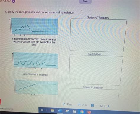
Table of Contents
- Classify The Myograms Based On Frequency Of Stimulation
- Table of Contents
- Classifying Myograms Based on Frequency of Stimulation
- Single Stimulus: The Twitch Contraction
- Latent Period: The Silent Phase
- Contraction Phase: The Rise of Tension
- Relaxation Phase: The Fall of Tension
- Low-Frequency Stimulation: Incomplete Tetanus
- High-Frequency Stimulation: Complete Tetanus
- Treppe: The Staircase Phenomenon
- Factors Affecting Myogram Classification
- Muscle Fiber Type:
- Muscle Temperature:
- Muscle Fatigue:
- Ionic Concentrations:
- Neuromuscular Junction Function:
- Clinical Significance of Myogram Classification
- Conclusion
- Latest Posts
- Latest Posts
- Related Post
Classifying Myograms Based on Frequency of Stimulation
Understanding muscle contractions is crucial in various fields, from physiology and sports science to rehabilitation and clinical diagnosis. Electromyography (EMG), the technique of recording the electrical activity of muscles, plays a pivotal role in this understanding. The resulting graphical representation of muscle activity is known as a myogram. A key factor influencing the appearance of a myogram is the frequency of stimulation applied to the muscle. This article will delve into the different classifications of myograms based on the frequency of stimulation, explaining the underlying physiological mechanisms and the characteristic features of each type.
Single Stimulus: The Twitch Contraction
The simplest form of muscle contraction is the twitch contraction, resulting from a single electrical stimulus. This myogram demonstrates a characteristic three-phase response:
Latent Period: The Silent Phase
The initial phase, the latent period, is a brief delay (typically 2 milliseconds) between the stimulus application and the onset of muscle contraction. During this time, the excitation-contraction coupling process is underway. This includes the depolarization of the muscle fiber membrane, the release of calcium ions from the sarcoplasmic reticulum, and the subsequent formation of cross-bridges between actin and myosin filaments. While no visible contraction occurs, significant intracellular events are preparing the muscle for action.
Contraction Phase: The Rise of Tension
Following the latent period, the contraction phase begins. This is characterized by a rapid increase in muscle tension as cross-bridges repeatedly cycle, leading to the sliding of actin and myosin filaments and the shortening of the sarcomeres. The peak of tension marks the end of this phase. The duration of the contraction phase is influenced by factors such as the muscle fiber type and temperature.
Relaxation Phase: The Fall of Tension
Finally, the relaxation phase sees a gradual decrease in muscle tension as calcium ions are actively pumped back into the sarcoplasmic reticulum, breaking the cross-bridges and allowing the muscle fibers to return to their resting length. This phase is generally slower than the contraction phase. The time taken for complete relaxation is also dependent on various factors including muscle type and temperature. A single twitch contraction provides a baseline for understanding more complex patterns of muscle activity generated by increased stimulation frequencies.
Low-Frequency Stimulation: Incomplete Tetanus
When a muscle is stimulated repeatedly at low frequencies (below the fusion frequency), the myogram shows a series of individual twitches separated by periods of relaxation. This is known as incomplete tetanus or unfused tetanus. Each subsequent stimulus triggers a new twitch before the muscle has fully relaxed from the previous one. The result is a series of increasingly stronger contractions, each building upon the previous one. This phenomenon is known as wave summation, where the additive effect of multiple stimuli results in progressively greater tension. The force generated increases because the calcium concentration in the cytosol remains elevated, as the rate of calcium removal is not fast enough to compensate for the repeated calcium release triggered by successive stimuli. This incomplete tetanus pattern is common during everyday activities involving rhythmic movements.
High-Frequency Stimulation: Complete Tetanus
As the frequency of stimulation increases beyond a critical point – the fusion frequency, the individual twitches fuse together to form a smooth, sustained contraction known as complete tetanus or fused tetanus. At this frequency, the muscle doesn't have time to relax between stimuli, resulting in a maximal, sustained contraction. The force generated is significantly higher than during incomplete tetanus because the calcium levels in the cytosol remain sufficiently high to maintain a maximal number of active cross-bridges. The sustained contraction appears as a smooth line on the myogram, highlighting the complete absence of relaxation phases between stimuli. This type of contraction is essential for activities requiring sustained force generation, such as maintaining posture or gripping an object firmly. The precise fusion frequency is dependent on the specific muscle involved and other physiological factors.
Treppe: The Staircase Phenomenon
In some circumstances, repeated low-frequency stimulation can lead to a phenomenon known as treppe, also referred to as the staircase effect. In this case, instead of a simple summation of twitches as seen in incomplete tetanus, the initial twitches are progressively stronger, reaching a plateau before eventually stabilizing. This increase in tension is not due to summation but rather to an increasing efficiency in excitation-contraction coupling. This is thought to be caused by a gradual increase in intracellular calcium concentration with repeated stimulation, leading to a greater number of active cross-bridges. Treppe is most noticeable after a period of rest and diminishes with continued stimulation. It is less frequently observed than the other myogram patterns described above.
Factors Affecting Myogram Classification
The classification of myograms based on stimulation frequency isn't solely determined by the frequency itself. Several other factors play significant roles:
Muscle Fiber Type:
The type of muscle fibers present within the muscle significantly impacts the myogram. Fast-twitch fibers contract and relax more rapidly than slow-twitch fibers, resulting in different responses to the same stimulation frequency. Fast-twitch fibers will exhibit a faster twitch contraction and reach tetanus at higher frequencies compared to slow-twitch fibers.
Muscle Temperature:
Temperature significantly affects the rate of enzyme activity, particularly those involved in excitation-contraction coupling and calcium handling. Increased temperature accelerates these processes, leading to faster contraction and relaxation times, thus impacting the appearance of the myogram. This means that the fusion frequency for complete tetanus will be altered by temperature changes.
Muscle Fatigue:
Prolonged stimulation can lead to muscle fatigue, where the ability of the muscle to generate force diminishes over time. Fatigue will alter the myogram, reducing the amplitude and duration of both twitch contractions and tetanus. The extent of fatigue-induced changes is dependent on the intensity and duration of stimulation, as well as the metabolic capabilities of the muscle fibers involved.
Ionic Concentrations:
The concentrations of ions such as calcium, sodium, and potassium are crucial for muscle excitation and contraction. Alterations in these ionic concentrations can significantly impact muscle function and thus the characteristics of the myogram. For example, hypocalcemia can weaken muscle contraction, affecting both the amplitude and fusion frequency.
Neuromuscular Junction Function:
The neuromuscular junction is the site where the motor neuron synapses with the muscle fiber. Any impairment in neuromuscular transmission will affect the muscle response to stimulation, altering the appearance of the myogram. Diseases such as myasthenia gravis can significantly alter the myogram response.
Clinical Significance of Myogram Classification
The ability to classify myograms based on stimulation frequency has significant clinical implications:
- Diagnosing Neuromuscular Disorders: Abnormal myograms can indicate underlying neuromuscular disorders. For example, myasthenia gravis can cause a reduction in the amplitude of muscle contractions and a failure to achieve complete tetanus.
- Assessing Muscle Function: Myograms can be used to assess the functional capacity of muscles, helping to monitor recovery after injury or disease. Changes in twitch characteristics or the ability to achieve complete tetanus can reflect improvements or deterioration in muscle function.
- Evaluating the Effects of Therapeutic Interventions: Myograms can be used to evaluate the effectiveness of therapeutic interventions, such as electrical stimulation or pharmacological treatments, on muscle function.
- Monitoring Muscle Fatigue: Changes in myograms during prolonged stimulation can help to monitor the development of muscle fatigue, providing valuable insights into the limits of human performance.
Conclusion
The classification of myograms based on stimulation frequency provides a powerful tool for understanding the intricacies of muscle contraction. By analyzing the characteristics of different myogram patterns, such as twitch contractions, incomplete tetanus, and complete tetanus, we can gain valuable insights into the underlying physiological mechanisms and their impact on muscle performance. Furthermore, the ability to interpret myograms holds significant clinical value in diagnosing and monitoring neuromuscular disorders, assessing muscle function, and evaluating the effectiveness of therapeutic interventions. A deeper understanding of this technique empowers healthcare professionals and researchers to enhance patient care and advance scientific knowledge. The intricate interplay of stimulation frequency, muscle fiber type, temperature, fatigue, and ionic concentrations underscores the complexity of muscle physiology and the importance of considering these factors when interpreting myograms.
Latest Posts
Latest Posts
-
Match The Neuroglial Cell With Its Correct Function
Mar 30, 2025
-
Health Economics And Policy James Henderson Cite Apa
Mar 30, 2025
-
Which Of The Following Statements About Cycloaddition Reactions Is True
Mar 30, 2025
-
Elisa Graduated From College With A Double Major
Mar 30, 2025
-
The Inflation Rate Is Defined As The
Mar 30, 2025
Related Post
Thank you for visiting our website which covers about Classify The Myograms Based On Frequency Of Stimulation . We hope the information provided has been useful to you. Feel free to contact us if you have any questions or need further assistance. See you next time and don't miss to bookmark.
