Art-labeling Activity Anatomy And Histology Of The Adrenal Gland
Holbox
Mar 27, 2025 · 6 min read
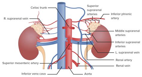
Table of Contents
- Art-labeling Activity Anatomy And Histology Of The Adrenal Gland
- Table of Contents
- Art-Labeling Activity: Anatomy and Histology of the Adrenal Gland
- I. Adrenal Gland: An Overview
- A. Size and Location
- B. Blood Supply
- II. Anatomical Art Labeling: External Features
- III. Histological Art Labeling: Microscopic Structure
- A. Adrenal Cortex
- B. Adrenal Medulla
- IV. Advanced Art Labeling Considerations
- V. Creating Engaging and Informative Illustrations
- VI. Application in Different Contexts
- VII. Conclusion
- Latest Posts
- Latest Posts
- Related Post
Art-Labeling Activity: Anatomy and Histology of the Adrenal Gland
The adrenal gland, a small yet vital organ nestled atop each kidney, plays a crucial role in regulating various bodily functions. Understanding its intricate anatomy and histology is paramount for accurate art labeling and a deeper comprehension of its physiological significance. This article delves into the detailed structure of the adrenal gland, providing a comprehensive guide for creating precise and informative anatomical illustrations and diagrams.
I. Adrenal Gland: An Overview
The adrenal glands, also known as suprarenal glands, are paired endocrine glands that produce a variety of hormones essential for maintaining homeostasis. Their location, immediately superior to the kidneys, contributes to their anatomical significance. Each gland is roughly pyramidal in shape, with a distinctive inner medulla and outer cortex. These two regions are functionally and histologically distinct, each contributing a unique set of hormones to the body's endocrine system.
A. Size and Location
The adrenal glands are relatively small organs, each typically measuring around 3-5 centimeters in length, 2-3 centimeters in width, and 1 centimeter in thickness. Their weight varies, typically ranging from 3 to 5 grams in adults. Their precise location, superior to the kidneys and embedded in the perirenal fat, protects them while maintaining close proximity to the renal vasculature. This location is crucial for their hormonal influence and their efficient delivery of hormones into the bloodstream.
B. Blood Supply
The rich vascularization of the adrenal glands is vital for their hormonal function. The adrenal glands receive a substantial blood supply from three major arteries:
- Superior suprarenal arteries: Branching from the inferior phrenic arteries.
- Middle suprarenal arteries: Branching directly from the abdominal aorta.
- Inferior suprarenal arteries: Branching from the renal arteries.
This extensive network ensures the rapid delivery of hormones into the systemic circulation, allowing for swift physiological responses. The venous drainage is primarily via the suprarenal veins, which eventually drain into the inferior vena cava.
II. Anatomical Art Labeling: External Features
When creating an art label for the adrenal gland, start by depicting its overall shape and location relative to the kidney. Accuracy is paramount. Include labels for:
- Adrenal Gland (Suprarenal Gland): This should be a prominent label, clearly identifying the entire organ.
- Right Adrenal Gland: Label the right gland specifically, highlighting its somewhat triangular shape.
- Left Adrenal Gland: Label the left gland, noting its slightly more crescent-shaped morphology.
- Kidney (Right & Left): Clearly show the relationship between the adrenal gland and the kidney, emphasizing the superior positioning.
- Inferior Vena Cava: Include this label, especially when illustrating the venous drainage of the right adrenal gland.
- Renal Artery: Indicate the location of the renal artery, highlighting its proximity to the inferior suprarenal artery.
- Abdominal Aorta: This label is crucial for demonstrating the origin of the middle suprarenal artery.
- Inferior Phrenic Artery: Show the origin of the superior suprarenal arteries.
- Perirenal Fat: Illustrate the surrounding fatty tissue, providing context to the gland's protective environment.
III. Histological Art Labeling: Microscopic Structure
The microscopic anatomy of the adrenal gland reveals a distinct zonal arrangement crucial to its endocrine function. Precise labeling of histological sections is vital.
A. Adrenal Cortex
The adrenal cortex, the outer layer, constitutes the majority of the gland's mass (about 80-90%). It is further subdivided into three distinct zones, each with unique histological characteristics and hormone production:
-
Zona Glomerulosa: This outermost zone is characterized by densely packed, rounded cells arranged in clusters or arches. Its cells are relatively small and contain numerous lipid droplets. Label this zone clearly and indicate its primary hormone product: mineralocorticoids (primarily aldosterone). Aldosterone's role in regulating sodium and potassium balance should be mentioned in any accompanying text.
-
Zona Fasciculata: This middle zone is the widest of the three cortical zones and is characterized by long, parallel cords of cells arranged in columns. These cells are larger than those in the zona glomerulosa and are filled with abundant lipid droplets. Label this zone and indicate its production of glucocorticoids (primarily cortisol). Cortisol's role in stress response, glucose metabolism, and immune regulation should be described in accompanying notes.
-
Zona Reticularis: This innermost zone of the cortex is characterized by a network of branching cords of cells. The cells are smaller and less lipid-rich than those in the zona fasciculata. Label this zone and note its production of adrenal androgens (e.g., DHEA, androstenedione). The roles of these hormones in sexual development and other metabolic processes should be clearly explained.
B. Adrenal Medulla
The adrenal medulla, the inner portion of the adrenal gland, is considerably smaller than the cortex. It is composed of chromaffin cells, which are modified postganglionic sympathetic neurons that secrete catecholamines (epinephrine and norepinephrine). Clearly label the medulla and highlight the chromaffin cells. Detailed labeling could include:
- Chromaffin Cells: Identify these specialized cells responsible for catecholamine synthesis and release.
- Blood Vessels: Show the rich vascular network supplying the medulla, facilitating rapid hormone release.
- Nerve Fibers: Indicate the presence of preganglionic sympathetic nerve fibers, which innervate the chromaffin cells.
- Epinephrine (Adrenaline): Clearly label this hormone and describe its role in the "fight-or-flight" response.
- Norepinephrine (Noradrenaline): Similarly, label norepinephrine and its role in regulating blood pressure and other sympathetic functions.
IV. Advanced Art Labeling Considerations
For more advanced anatomical illustrations and histological diagrams, consider including these details:
- Connective Tissue: Show the presence of connective tissue capsules and septa that support the adrenal gland's structure.
- Lymphatic Vessels: Illustrate the presence of lymphatic vessels, contributing to immune surveillance of the gland.
- Histological Stains: Specify the type of stain used in the histological section (e.g., H&E stain, special stains for lipid visualization).
- Magnification: Indicate the magnification level of the microscopic image.
- Key: Include a clear key or legend for all labels within the illustration.
V. Creating Engaging and Informative Illustrations
The effectiveness of your art labeling hinges not only on accuracy but also on clarity and visual appeal. Consider these points:
- Use Clear and Concise Labels: Avoid overly long or technical terms; prioritize clarity and readability.
- Use Different Colors and Font Sizes: Differentiate structures and labels for better visual distinction.
- Maintain a Consistent Style: Maintain a unified aesthetic throughout the illustration or diagram.
- Use Arrows and Lines to Connect Labels: Guide the viewer's eye to specific structures.
- Contextual Information: Include brief descriptive text explaining the functions of the labeled structures.
VI. Application in Different Contexts
Accurate art labeling of the adrenal gland is essential in several contexts:
- Medical Education: For teaching medical students and residents about endocrinology and anatomy.
- Medical Textbooks and Journals: To illustrate anatomical and histological details in publications.
- Patient Education Materials: To help patients understand their condition in cases involving adrenal gland disorders.
- Research Presentations: To visualize research findings related to adrenal gland structure and function.
VII. Conclusion
Creating accurate and informative art labels for the adrenal gland requires a thorough understanding of its complex anatomy and histology. By following the guidelines outlined in this article, you can produce visually appealing and scientifically accurate illustrations that effectively communicate the intricacies of this vital endocrine organ. Remember, the goal is to create images that are not only visually engaging but also enhance understanding and promote accurate knowledge dissemination. The application of these principles extends beyond academic settings, benefiting medical professionals, researchers, and patients alike. Through precise and detailed labeling, you contribute to a clearer and more comprehensive understanding of this crucial organ and its vital role in human physiology.
Latest Posts
Latest Posts
-
Juanjo Y Manuel No Encuentran El Puesto De Gafas
Mar 31, 2025
-
Which Of The Following Is True Of Services
Mar 31, 2025
-
Which Of The Following Elements Is Present In This Image
Mar 31, 2025
-
You Have Important Time Sensitive Information
Mar 31, 2025
-
Liquid Sodium Is Being Considered As An Engine Coolant
Mar 31, 2025
Related Post
Thank you for visiting our website which covers about Art-labeling Activity Anatomy And Histology Of The Adrenal Gland . We hope the information provided has been useful to you. Feel free to contact us if you have any questions or need further assistance. See you next time and don't miss to bookmark.
